A 63-year-old man says the 20-year-old rash on his face first appeared one summer. Although it slackens a bit each winter, it flares up again when the weather warms—despite a bevy of OTC and prescription topical treatments.
The patient has consulted many providers, including several dermatologists, who have diagnosed the butterfly rash of lupus. But blood tests failed to bear out that theory, and no one has ever biopsied it.
The patient spent many years working in the sun with minimal to no protection. He denies fever, malaise, joint pain, or other illness. He denies having a similar rash elsewhere on his body.
EXAMINATION
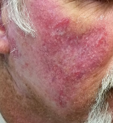
A symmetrical, strikingly red, extensive rash covers most of both sides of the patient’s face. There is epidermal scaling and roughness and large areas of obvious follicular enlargement, atrophy, and telangiectasias.
Punch biopsy shows a multitude of changes: atrophic epithelium, basal vacuolar changes, an intense dermal lymphocytic infiltrate, liquefaction degeneration, and apoptotic keratinocytes. Compact orthokeratosis is noted on the surface, and increased mucin formation in the dermis.
What is the diagnosis? DISCUSSION
These skin changes, in conjunction with the biopsy findings, are consistent with a diagnosis of discoid lupus erythematosus (DLE). The vast majority of affected patients do not have systemic lupus erythematosus (SLE), although nearly 17% eventually progress to it. This patient’s seronegative status had puzzled his previous providers—a common mistake that frequently delays diagnosis and treatment.
DLE is the most common type of chronic cutaneous lupus, occurring in about 17 to 48 of every 100,000 people in the general population. It affects far more women than men, mostly those ages 20 to 40. More blacks than whites are affected by DLE, which is thought to be a polygenic autoimmune disease linked to various human leukocyte antigen groups.
DLE affects sun-exposed areas, including the scalp (where it often goes undiagnosed, leading to scarring alopecia). It can also occur in the mouth, manifesting as annular eroded areas. In this patient’s case, the combination of UV exposure and apparent genetic predisposition caused his malady.
Following workup to rule out systemic disease, the patient was started on hydroxychloroquine HCL (200 mg bid) and given strict instructions to protect himself from the sun. This should yield considerable improvement, although the scarring on large sections of his face (eg, the posterior cheeks) will be permanent. He will also be monitored for possible progression to SLE.
TAKE-HOME LEARNING POINTS
- Discoid lupus erythematosus (DLE) is a form of chronic cutaneous lupus caused by overexposure to the sun in a genetically susceptible individual.
- Most DLE patients never develop systemic lupus, though it’s far from unknown (17%).
- DLE, an autoimmune disease, is far more common in women (ages 20 – 40) than in men.
- Diagnosis is often made clinically, but biopsy reveals characteristic findings that confirm the disease.
- Besides sun protection, DLE is treated with the oral antimalarial hydroxychloroquine HCL (200 mg bid) and topical steroids as needed.