DermaDiagnosis

Sticking His Neck Out Leads to Diagnosis

Author and Disclosure Information

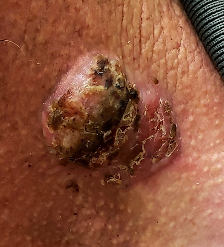
Neck lesion

For 5 years, a lesion has been slowly growing on this 50-year-old man’s neck. He has seen several primary care providers who had provided different diagnoses, including wart, seborrheic keratosis, and keratoacanthoma. Throughout these visits, the only treatment he was offered was cryotherapy, but this was not effective at clearing the lesion. One family member convinced him it was time to try a dermatology provider.

In his lifetime, the patient has been exposed to a great deal of UV light. He has had several basal cell and squamous cell carcinomas removed from his face and arms.

He claims to be in otherwise good health, but further questioning reveals a > 40 pack-year history of smoking. He also has a recent diagnosis of early chronic obstructive pulmonary disease.

Physical examination of the lesion reveals a 4-x-3.5-cm sessile mass located on the left lateral neck. Its surface is rough and irregular, but there is no break in the skin.

On palpation, the lesion is quite firm and only partially mobile. No tenderness is detected. There are no palpable nodes in the region. There is evidence of advanced chronic sun damage on all sun-exposed areas, but especially on the head and neck. The rest of skin has no lesions.

The differential for this lesion includes

squamous cell carcinoma

basal cell carcinoma

keratoacanthoma

all of the above

ANSWER

The correct answer is all of the above (choice “d”).

DISCUSSION

For this patient, there could have been even more items in the differential, including melanoma, Merkel cell carcinoma, or even metastatic (from lung, colon, etc) origin. Dermatofibroma sarcoma protuberans is another possibility because it is rarely aggressive, though it is seldom as exophytic as this lesion. However, the prolonged, indolent course of the patient’s lesion was more consistent with the 3 choices listed. The overarching point is this: Why guess when the diagnosis is easily obtained by biopsy?

Biopsy of the lesion was ordered at the patient's first visit to dermatology. It revealed an invasive basal cell carcinoma (BCC), which almost never metastasizes. BCC requires extensive surgical removal and closure.

TREATMENT

The patient’s lesion needed a total excision with margins. Its size, location, and longevity called for the Mohs technique to assure clear margins and optimal closure.

Because of the findings and the patient’s history, he would also need to see dermatology every 6 months because he is a prime candidate for developing other skin cancers.

Recommended Reading

Beware a pair of dermatologic emergencies in children
Clinician Reviews
Preadolescent acne: Management from birth requires increasing vigilance
Clinician Reviews
Food allergy testing for eczema in kids varies by specialty
Clinician Reviews
Shortcomings identified in study of acne videos on TikTok
Clinician Reviews
More severe AD correlates with worse sleep health and attention problems in children
Clinician Reviews
Light-based technologies emerging as promising acne treatments
Clinician Reviews
Swedish registry study finds atopic dermatitis significantly associated with autoimmune diseases
Clinician Reviews
Expert picks top pediatric dermatology studies of 2020
Clinician Reviews
Avoiding atopic dermatitis triggers easier said than done
Clinician Reviews
Baseline body surface area may drive optimal baricitinib responses
Clinician Reviews