DermaDiagnosis

Why Size (of Your Differential) Matters

Author and Disclosure Information

Itchy lesion

A 50-year-old man is astonished when his “fungal infection” fails to respond to an unknown OTC topical cream a friend advised him to use.

For several weeks, he’s been plagued by the slightly itchy lesions that appeared on his leg without known cause. His friend assured him that the shape and configuration of the lesions could only represent one thing: “ringworm,” that is, fungal infection.

However, in clinic, the patient denies any contact with animals or children and reports that his job is inside only, never involving the great outdoors. He has felt fine throughout the lesions’ tenure, going so far as to say he is “in perfect health.” He has been in a mutually monogamous relationship for many years.

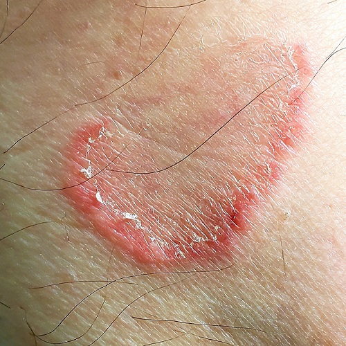
The lesions (4 in total) are located on the medial aspect of the left leg, extending into the popliteal area. At first glance, they appear to be peripherally scaly, pink, and annular. Close inspection reveals that most of the scaling is not on the outer edge; it is confined to the inside of the border, a phenomenon termed trailing or centripetal scaling. The lesions all show an arciform morphology in the shape of a “C.”

KOH examination of the scale shows no fungal elements. Shave biopsy reveals a dense perivascular lymphocytic infiltrate, moderate parakeratosis, and perhaps most importantly, no fungal elements in the stratum corneum.

There are no palpable lymph nodes in the groin on the affected side.

What items could be in the differential for this lesion?

Psoriasis

Nummular eczema

Erythema annulare centrifugum

Secondary syphilis

A variety of paraneoplastic eruptions

All of the above

ANSWER

The correct answer is all of the above (choice “f”).

DISCUSSION

The most likely diagnostic explanation for this patient’s presentation is erythema annulare centrifugum (EAC; choice “c”). However, this diagnosis is often difficult to establish, in part because of the broad differential and also because the very existence of EAC is far from well established.

The overwhelming consensus is that EAC represents a hypersensitivity reaction to an unknown antigen. It can be triggered by a wide variety of micro-organisms, stress, or even pregnancy.

In this case, there was no clinical or historical reason to suspect an underlying cancer, Lyme disease, or lupus, nor did the biopsy results suggest any of these.

The key takeaway here is to urge providers to avoid jumping onto a diagnostic bandwagon before considering a wider differential. Indeed, size matters when it relates to the length of one’s differential diagnosis list. If you don’t consider it, you can’t diagnose it.

TREATMENT

Fortunately, EAC nearly always resolves, with or without treatment. This patient received reassurance as such but was scheduled to return for a check of his lesions in 2 months. At that point, they had resolved.

Recommended Reading

Brown plaque on the arm
Clinician Reviews
Making the Rounds to Diagnosis
Clinician Reviews
Numerous large nodules on scalp
Clinician Reviews
Scaly patches on hand and feet
Clinician Reviews
All’s Well That Ends Swell(ing)
Clinician Reviews
Scaly beard rash
Clinician Reviews
Foot rash and joint pain
Clinician Reviews
New analysis puts U.S. psoriasis prevalence at 3%
Clinician Reviews
Study eyes impact of isotretinoin on triglycerides, other lab measures
Clinician Reviews
Stop using Neutrogena and Aveeno spray sunscreen, J&J warns
Clinician Reviews