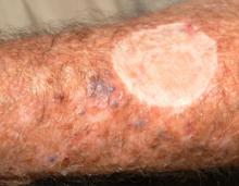
A 73-year-old man self-refers to dermatology 18 months after a melanoma was diagnosed and removed from his forearm. Following that discovery, he was referred to a surgeon, who performed a wide excision (the defect from which was closed with a graft) and who went on to do lymph node dissection in the ipsilateral axilla. No positive nodes were found.
The wounds from these procedures are long since healed, and the patient has been doing well. That is, until recently, when he noticed some new lesions developing around the graft site.
EXAMINATION
About 15 to 20 firm, blue-black papules and nodules surround the periphery of the graft site on the patient’s forearm. Some extend out as far as 10 cm, though most are within 3 cm. Obviously intradermal, these lesions display no surface change at all. Punch biopsy confirms the suspicion that these represent satellite metastasis of the patient’s original melanoma, which itself had been more than 3 mm thick.
Fortunately, no nodes are palpable in the axilla, and no evidence of metastasis is found on physical examination, blood work, and PET scan.
DISCUSSION
The image accompanying this case is pregnant with information—some obvious, some less so. For example, the multiple blue-black nodules can easily be seen surrounding the graft site and were just as easily palpated.
Even ignoring those lesions momentarily, a look at the surrounding skin offers a veritable textbook of germane information. The collective term for the skin changes on the patient’s arms is dermatoheliosis, or sun-damaged skin. But that term comprises a number of specific changes, all of which have names and significance.
The casual observer might simply chalk these changes up to age, but for medical providers, more specifics are in order: The sun has thinned the patient’s skin remarkably, hence the term solar atrophy. His dorsal forearms are greatly discolored as well, changes we call poikiloderma. Numerous telangiectasias (also sun-caused) can be seen on his dorsal forearms. These changes are especially appreciated when the dorsal forearm skin is compared to the extensor forearms, which receive relatively little sun exposure.
The point? This patient had every reason to develop a melanoma, making any odd lesion on his skin suspicious. It also means his chances of developing a new primary melanoma are all too real, even if he survives the current one.
As one might imagine, this local recurrence of his melanoma is not a good sign at all. Strictly speaking, it is a form of metastasis—but until it reaches lymph nodes or organs, it only suggests that possibility.
Treatment choices are limited for his melanoma, but include limb perfusion, chemotherapy, and surgery. The truth is, his prognosis is poor. His case emphasizes the need for prevention and early diagnosis, the latter greatly aided by the recognition of patients at risk by virtue of having fair, sun-damaged skin.
As often happens in cases like this, there is a ripple effect as the news of his situation reaches family and friends, whose own skin becomes the subject of attention. In such cases, it’s not unusual for the whole family to then be seen in dermatology over the succeeding months—not only to be examined, but also hopefully educated in terms of prevention and recognition.
TAKE-HOME LEARNING POINTS
• Local recurrence of melanoma is common, especially with primary tumors that exceed 3 mm in thickness.
• UV overexposure has been established as the major contributor to development of melanoma.
• Melanoma is far more common in fair-skinned individuals than in those with darker skin; “fair” is defined as tolerating sun poorly, burning easily, and tanning poorly, if at all.
• Evidence of this excessive sun damage is called dermatoheliosis and consists of specific findings including solar atrophy, telangiectasias, and pigmentary alteration known as poikiloderma.
• The lack of effective treatment for metastatic melanoma underlines the necessity for prevention (protection from the sun) and early detection.

