DermaDiagnosis

These Old Lesions? She’s Had Them for Years …

Author and Disclosure Information

A 65-year-old woman is referred to dermatology with discoloration of her legs that started several weeks ago. Her family suggested it might be “blood clots,” although she has been taking warfarin since she was diagnosed with atrial fibrillation several months ago. Her dermatologic condition is basically asymptomatic, but the patient admits to scratching her legs, saying it’s “hard to leave them alone.” On further questioning, she reveals that she has had “rough places” on her legs for at least 20 years and volunteers that her sister had the same problem, which was diagnosed years ago as “fungal infection.” Both she and her sister spent a great deal of time in the sun as children, long before sunscreen was invented. The patient is otherwise fairly healthy. She takes medication for her lipids, as well as daily vitamins. Her atrial fibrillation is under control and requires no medications other than the warfarin. A great deal of focal discoloration is seen on both legs, circumferentially distributed from well below the knees to just above the ankles. Many of the lesions are brown macules, but more are purplish-red, annular, and scaly. On closer examination, these lesions—the ones the patient says she has had for decades—have a very fine, thready, scaly border that palpation reveals to be tough and adherent. They average about 2 cm in diameter. There are no such lesions noted elsewhere on the patient’s skin. There is, however, abundant evidence of excessive sun exposure, characterized by a multitude of solar lentigines, many fine wrinkles, and extremely thin arm skin.

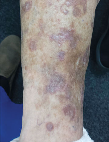
The most likely diagnosis for the leg lesions is

a) Disseminated superficial actinic porokeratosis

b) Stasis dermatitis

c) Seborrheic keratoses

d) Nummular eczema

ANSWER

The correct answer is disseminated superficial actinic porokeratosis (DSAP; choice “a”). This condition, caused by an inherited defect of the SART3 gene, is seen mostly on the sun-exposed skin of middle-aged women.

Stasis dermatitis (choice “b”) can cause a number of skin changes, but not the discrete annular lesions seen with DSAP.

Seborrheic keratoses (choice “c”) are common on the legs. However, they don’t display this same morphology.

Nummular eczema (choice “d”) presents with annular papulosquamous lesions (as opposed to the fixed lesions seen with DSAP), often on the legs and lower trunk, but without the thready circumferential scaly border.

Continue reading for Joe Monroe's discussion...

Pages

Recommended Reading

Hair Loss at a Very Young Age
Clinician Reviews
Man, 45, With Greasy Rash and Deformed Nails
Clinician Reviews
Being “Puzzled” as the First Step to Diagnosis
Clinician Reviews
FDA Approves Two-drug Combination for Advanced Melanoma
Clinician Reviews
A 10-Year Wait for Diagnosis
Clinician Reviews
Man Seeks Treatment for Periodic “Eruptions”
Clinician Reviews
If Not Fungal Infection, Then What?
Clinician Reviews
How to Manage a Keloid
Clinician Reviews
The Art and Science of Detecting Allergic Contact Dermatitis
Clinician Reviews
Case Studies in Toxicology: The Acclaimed Zombie-Apocalypse Drug—Is it Just an Illusion?
Clinician Reviews