DermaDiagnosis

Small, Painful Blisters Erupt in Patient's Groin Area

Author and Disclosure Information

What is causing this eruption of painful blisters?

The painful blisters are accompanied by fatigue and myalgia.


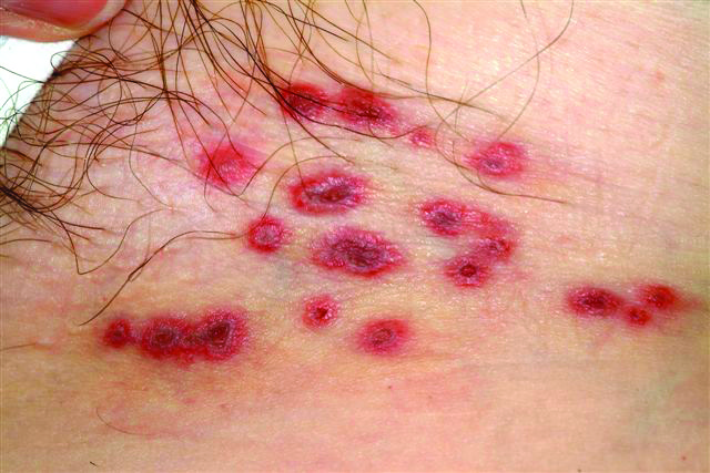
A 60-year-old woman presents with a symptomatic eruption in the left groin that began a week ago, when she first noticed a slight tingling sensation in that area. Within 48 hours, small blisters appeared, clustered in the crural fold. These became slightly painful, with a curious lancinating type of pain. The patient also began to experience fatigue and myalgia. She sought care when her lesions became hemorrhagic and was subsequently referred to dermatology. Additional history reveals that in the past month she has experienced low back pain on the left side, radiating into the groin. In response, she started to use moist hot packs on the area. The onset of her symptoms has coincided with another stressor, her husband’s job loss. She denies the use of any immunosuppressive medications and is well in other respects. Examination shows a grouped collection of targetoid hemorrhagic flaccid low blisters, from 1 to 2 cm in diameter, confined to the left crural fold. No underlying induration is felt, but there is palpable adenopathy confined to that area.

Based on the information provided, the most likely diagnostic explanation for her complaint is:

a) Yeast infection

b) Methicillin-resistant Staphylococcus aureus (MRSA)

c) Herpes simplex

d) Herpes zoster

ANSWER
The correct answer is herpes zoster (choice “d”; see discussion). Yeast infections (choice “a”) would be quite unlikely to present with hemorrhagic discrete lesions and far more likely to pre­sent with a confluent rash. MRSA (choice “b”) can present in a wide variety of forms but is unlikely to form discrete blisters and would almost certainly involve induration in the area. Without a definitive viral culture, herpes simplex (choice “c”) could not be ruled out in this case, since it presents in a similar manner. But given the patient’s age and the morphology and location of the lesions, zoster was far more likely. See the discussion for further differentiating factors.

DISCUSSION/TREATMENT
The appearance of intralesional hemorrhage in an inflammatory lesion bespeaks a deeper, more vigorous process, since vasculature ends just below the dermoepidermal junction. This occurs infrequently enough with herpes zoster to obscure the diagnosis. Ironically, herpes simplex almost never exhibits this phenomenon, making it useful as a differentiating feature.

In this patient’s case, it seems reasonable to blame the local application of heat as a potential trigger. My experience is that stress is also a factor, though the literature does not bear out that theory.

Recommended Reading

Systemic Scleroderma: The Truth Beneath a "Skin Disease"
Clinician Reviews
Malpractice Chronicle
Clinician Reviews
Malpractice Chronicle
Clinician Reviews
Malpractice Chronicle
Clinician Reviews
Dark Blue Facial Lesion Worries Mother of Young Boy
Clinician Reviews
What Is This Long-Standing Loss of Pigment?
Clinician Reviews
Skin Changes on Hands Lead to Daily Living Problems
Clinician Reviews
Two Different Diagnoses, But One Location
Clinician Reviews
If it’s not acne, warts, or molluscum ­contagiosum, what is this condition?
Clinician Reviews