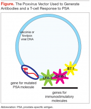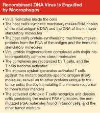
This recombinant viral vector is propagated in tissue culture, purified, and administered to the patient subcutaneously. The virus replicates for a short period in the host, expressing the 3 immunostimulatory molecules and the mutated PSA molecule protein product, which differs by 1 amino acid from that expressed by the patient’s tumor. The vaccinia virus and the mutant protein are recognized as foreign by the patient’s immune system, whereas the 3 immunostimulatory molecules are not, as they are the same as the host’s immunostimulatory molecules.
Subsequent boosts, administered over about 5 months, are with a fowlpox virus vector (which does not replicate in the patient) containing the same 4 genes that had been inserted into the vaccinia virus. This strategy enhances the immune response to the mutant tumor product and ensures that the host is not generating a response to the vaccinia virus. The mutant tumor product is sufficiently similar to the patient’s native tumor product that the immune system mounts a response to both proteins. The T cells generated in response to the mutant protein bind to both PSA and mutated PSA molecule proteins, and cells harboring these proteins are destroyed. 10
Whereas the T-cell response is important for the activity of the PROSTVAC vaccine, and there is no evidence for formation of anti-PSA antibodies, other vaccines exploit both B- and T-cell responses. 10,11 After a virus commandeers the host cell’s replication, transcription, and translation machinery, the macrophage breaks down the viral protein product (the mutated or immunogenic molecule) into small fragments. These fragments bind to major histocompatibility complex (MHC) class II molecules, which are produced by the macrophages. These complexes of antigen (virus) fragments with MHC class II molecules are transported to the surface of the macrophage, where the antigen is presented to lymphocytes. A receptor on a B lymphocyte adheres to the viral antigen and thus becomes an activated B cell, which divides and produces many copies. The B lymphocytes mature into plasma cells and release antibodies that adhere to the virus. A helper T-cell receptor can adhere to the antigen-MHC class II complex displayed on the B cell and activate the lymphocyte, causing the release of more and different cytokines. The cytokines stimulate the activated B cell to divide, producing functionally mature antibodies that recognize and attach to the virus. Macrophages can then engulf and destroy the virus, or the antibody-linked viruses can be excreted in the urine or stool. 12
When viruses (eg, the recombinant vaccinia virus of the PROSTVAC vaccine) infect cells and replicate, fragments of viral proteins become attached to MHC class I molecules (see Box). These complexes attach to the cell surface and are presented to cytotoxic T cells. The activated cytotoxic T cells divide and destroy cells harboring the virus or viral antigens. 12
Some of the B lymphocytes become memory B cells. Similarly, some of the T lymphocytes become memory T cells. These memory B cells respond and replicate rapidly on re-exposure to the virus or viral proteins to produce antigen-specific antibodies. 12


