Introduction
Direct visualization of the biliary ductal system is quickly gaining importance among gastroenterologists. Since the inception of cholangioscopy in the 1970s, the technology has progressed, allowing for ease of use, better visualization, and a growing number of indications. Conventional endoscopic retrograde cholangiopancreatography (ERCP) is successful for removal of bile duct stones (with success rates over 90%);1 however, its use in the evaluation of potential biliary neoplasia has been somewhat disappointing. The diagnostic yield of ERCP-guided biliary brushings can range from 30% to 40%.2-4 An alternative to ERCP-guided biliary brushings for biliary strictures is endoscopic ultrasound (EUS)-directed fine needle aspiration (FNA), but the reported sensitivity remains poor, ranging from 43% to 77% with negative predictive values of less than 30%.5-7 These results leave much to be desired for diagnostic yield.
The newest method of evaluating pancreaticobiliary pathology is with direct visualization using cholangioscopy. The advantages of this modality include the ability to obtain direct visualization as well as targeted biopsies of suspicious lesions. The first fiberoptic cholangioscope was introduced in 1965 and the first use of peroral cholangioscopy was reported in in the mid 1970s.8,9 Early models were limited by their delicacy, relative immmobility, lack of dedicated irrigation channel, and need for two endoscopists using a “mother baby” design. Fiberoptic single-operator cholangiopancreatoscopy (FSOCP) was first introduced in 2006 by Boston Scientific (Marlborough, MA).10 It was designed to address the previously stated shortcomings of the first-generation cholangioscopy devices. Since its introduction, it has gained worldwide popularity in the diagnosis and management of pancreaticobiliary pathology and complex biliary stones.The initial model employed a reusable fiber optic optical probe, a disposable cholangioscope access and delivery catheter, and disposable small-caliber biopsy forceps. The components can be introduced through a duodenoscope that has a minimum working channel diameter of 3.4 mm. The original FSOCP catheter is attached to the duodenoscope by a silastic belt just below the operating channel, allowing for single operator use. The access and delivery catheter has an outer diameter of 10 F and three separate ports: an optical port, two dedicated 0.6-mm irrigation channels, and a 1.2-mm accessory channel that accepts various accessories including the small-caliber biopsy forceps, electrohydraulic lithotripsy (EHL) fibers, or a holmium laser probe. The catheter has fourway tip deflection. The fiberoptic probe does have limitations, including its limited field of view, fragility of the fiber, and need for adjustment of the lens focus. Because of these limitations, a digital single-operator cholangioscope (DSOCP) was developed and introduced in 2014 (Boston Scientific, Marlborough, MA). In the DSOCP system, the light is generated by two independent light-emitting diodes and a complementary metal-oxide semiconductor digital camera chip. Improvements included a wider 120-degree field of view, dedicated irrigation and aspiration channels/connections, suction channel, and redesigned accessory channel. The cholangioscope is entirely disposable. The processor receives video signals from the catheter, processes the signals and outputs video images to an attached monitor. The newer digital-based platform has shown promising results, including higher diagnostic yield and shorter ERCP completion time when compared with similarly performed procedures using the fiberoptic-based platform.11Clinical indications
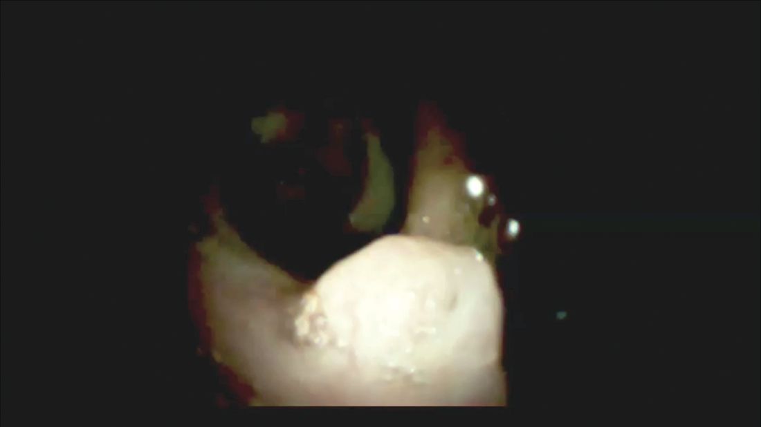
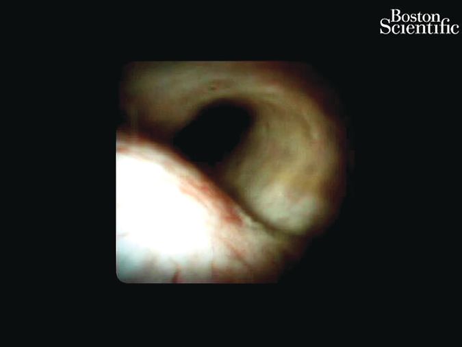
Direct visualization and biopsy of indeterminate biliary strictures has resulted in greatly improved diagnostic accuracy and collection of adequate tissue (Figures. 1,2). In a recent systematic review, the pooled sensitivity and specificity of cholangioscopy-guided biopsies in the diagnosis of malignant biliary strictures was 61% (95% confidence interval, 55%-65%) and 98% (95% CI, 96%-99%), respectively. Direct comparison of small-caliber direct biopsies with standard brushings and biopsies showed small-caliber direct biopsies having a sensitivity of 76.5% versus 5.8% and 29% with standard brushes and biopsies, respectively.12 The pooled sensitivity and specificity of six studies using cholangioscopy with targeted biopsies in the diagnosis of cholangiocarcinoma was 66.2% and 97.0%, respectively.12 Studies have shown that small-caliber forceps obtains tissue adequate for pathologic evaluation in 82%-97% of biopsy samples retrieved.13-17 Three prospective trials have evaluated the diagnostic accuracy of small-caliber forceps for indeterminate biliary lesions. The accuracy ranged from 72% to 85% with a sensitivity of 49%-82%, specificity of 82%-100%, positive predictive value of 100%, and negative predictive value of 69%-100%.15-17 The improved diagnostic accuracy of cholangioscopy for indeterminate biliary strictures stems from its direct visualization ability. Traditional sampling techniques (cytology brushings and fluoroscopically guided biopsies) are plagued by low sensitivity and negative predictive value caused by a relatively high false-positive rate.DSOCP appears to have improved accuracy over fiberoptic equipment. In a recent multicenter observational study in patients undergoing digital cholangioscopy, the guided biopsies resulted in adequate tissue for histologic evaluation in 98% of patients. In addition, the sensitivity and specificity of digital cholangioscope-guided biopsies for diagnosis of malignancy was 85% and 100%, respectively.11
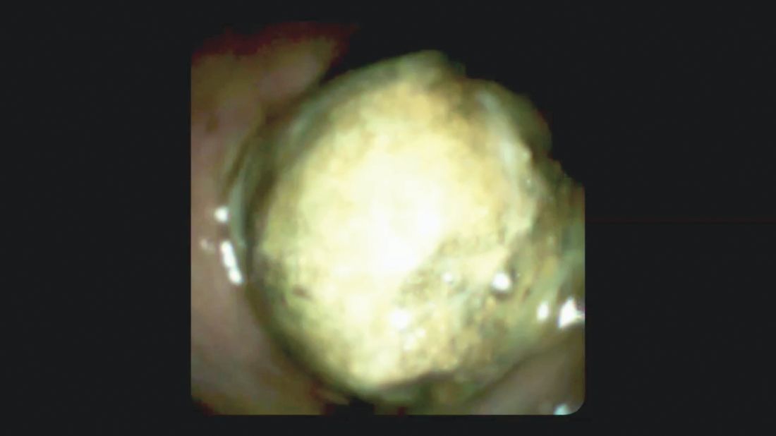
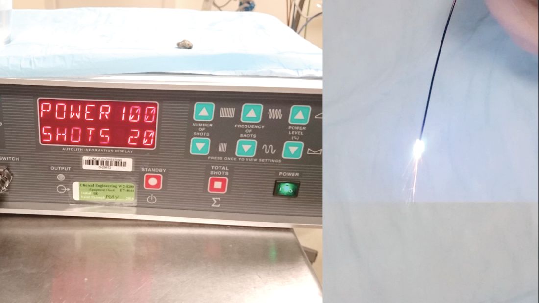
Conventional ERCP is successful in most cases of biliary stone extraction but, in 5%-10% of cases, stones can be difficult to remove because of size, location above strictures, or adherence to the bile duct wall18 (Figure 3). In addition, lithotripsy with standard fluoroscopic guidance can cause stone fragments to get lost. In one study, 29% of ERCP-lost gallstones were diagnosed by post-hoc cholangioscopy.19 A number of studies have documented a high success rate of FSOCP- or DSOCP-guided lithotripsy, ranging from 90% to 100% (13,14,16,20,21). In addition cholangioscopy can circumvent the need for mechanical lithotripsy. EHL is used for the majority of cases, but use of a holium laser has also been described.20,21 The dedicated irrigation channels on the FSOCP/DSOCP system give the ability to continuously fill the biliary system with fluid, which is required for EHL (Figures 4,5).Diagnostic pancreatoscopy has advantages in the diagnosis and future management of malignancies and intraductal papillary mucinous neoplasms (IPMNs). In addition, pancreatic duct stones can easily be managed with digital pancreatoscopy and lithotripsy (EHL or laser lithotripsy). A study that included 115 patients that were followed for at least 2 years showed that pancreatoscopy was able to diagnose 63% of pancreatic cancers, 80% of benign strictures, and 95% of intraductal papillary mucinous neoplasms based on visual appearances. The authors were able to discern neoplasia based on visual findings, including coarse or granular mucosa, protrusion, papillary tumor, and tumor vessel.22 In a similar study, patients with confirmed intraductal papilliary mucinous neoplasms (IPMN) underwent peroral pancreatoscopy and/or intraductal ultrasound preoperatively. The detected protruding lesions were classified into five groups: granular mucosa, fish-egg with or without vascular images, villous type, and vegetative type. The diagnostic accuracy of peroral pancreatoscopy in differentiating benign IPMN from malignant ones was 88% with a sensitivity and specificity of 100% and 71% in the main duct type, respectively, and sensitivities and specificities of 43% and 100% of branch type, respectively.23DSOCP also has therapeutic implications for other pancreatic diseases. Pancreatic duct obstruction can be caused by stones and strictures. A large multicenter study of 1,000 patients with chronic pancreatitis revealed obstruction of the main pancreatic duct (MPD) in 50%; with 32% being caused by strictures and stones, while 18% were due solely to stones.24 Currently accepted treatments for pancreaticolithiasis include extracorporeal shock wave lithotripsy, ERCP with stone clearance, and stenting or surgery (pancreaticojejunostomy) but these techniques have limitations and can incur morbidity.DSOCP has recently been evaluated as an alternative technique in treating MPD stones. In a recent study, Bekkali et al reviewed their 3-year experience of digital pancreatoscopy and EHL for pancreatic duct stones. Of the pancreatoscopy procedures performed, 7% were for pancreatic stones. All the patients had painful chronic pancreatitis, radiographic evidence of a dilated pancreatic duct, and MPD stone disease. Stone fragmentation and pancreatic duct decompression were achieved in 83% without complications. Two patients required two EHL procedures to achieve clearance. In the single patient with failed clearance, pancreatoscopy revealed the stone to be in adjacent parenchyma and not in the pancreatic duct. All patients with successful pancreatoscopy and EHL had pain relief and marked improvement during follow up.25Other less common diagnostic indications for DSOCP include evaluation of cystic lesions of the biliary tract, verifying clearance of bile duct stones, bile duct ischemia evaluation after liver transplantation, hemobilia evaluation, removal of a bile duct foreign body, and evaluation of bile duct involvement in the presence of an ampullary adenoma.3,14,15,20,26,27

 Dr. Sonnier, Dr. Mizrahi, and Dr. Pleskow
 Dr. Sonnier, Dr. Mizrahi, and Dr. Pleskow