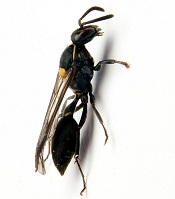
Polybia paulista
Photo by Mario Palma/
São Paulo State University
The wasp Polybia paulista protects itself from predators by producing venom known to contain a cancer-fighting toxin.
A study published in Biophysical Journal helps explain how the venom’s toxin, MP1 (Polybia-MP1), selectively kills cancer cells without harming normal cells.
MP1 interacts with lipids that are abnormally distributed on the surface of cancer cells, creating holes that facilitate the escape of molecules crucial for cell function.
“Cancer therapies that attack the lipid composition of the cell membrane would be an entirely new class of anticancer drugs,” said study author Paul Beales, PhD, of the University of Leeds in the UK.
MP1 is known to act against microbial pathogens by disrupting the bacterial cell membrane. The peptide has shown promise for treating cancers, as it can inhibit the growth of prostate and bladder cancer cells, as well as multi-drug resistant leukemic cells.
However, it has not been clear how MP1 selectively destroys cancer cells without harming normal cells. Dr Beales and his colleagues thought an explanation might lie in the unique properties of cancer cell membranes.
In healthy cell membranes, the phospholipids phosphatidylserine (PS) and phosphatidylethanolamine (PE) are located in the inner membrane leaflet facing the inside of the cell. But in cancer cells, PS and PE are embedded in the outer membrane leaflet facing the cell surroundings.
The researchers tested their theory by creating model membranes, some of which contained PE and/or PS, and exposing them to MP1. They used a wide range of imaging and biophysical techniques to characterize MP1’s destructive effects on the membranes.
The team found that PS increased the binding of MP1 to the membrane by a factor of 7 to 8. On the other hand, PE enhanced MP1’s ability to quickly disrupt the membrane, increasing the size of holes by a factor of 20 to 30.
“Formed in only seconds, these large pores are big enough to allow critical molecules such as RNA and proteins to easily escape cells,” said study author João Ruggiero Neto, of São Paulo State University in Brazil.
“The dramatic enhancement of the permeabilization induced by the peptide in the presence of PE and the dimensions of the pores in these membranes was surprising.”
In future studies, the researchers plan to alter MP1’s amino acid sequence to examine how the peptide’s structure relates to its function and further improve the peptide’s selectivity and potency for clinical purposes.
“Understanding the mechanism of action of this peptide will help in translational studies to further assess the potential for this peptide to be used in medicine,” Dr Beales said.
“As it has been shown to be selective to cancer cells and non-toxic to normal cells in the lab, this peptide has the potential to be safe, but further work would be required to prove that.”
