You never know what will happen when you look into a mouse’s eyes.
Twelve years ago, Dr. Lee Goldstein was investigating reactive oxygen species’ effect on the brain of a young Alzheimer’s model mouse. Holding the tiny creature in his hand, he carefully inserted a miniscule microdialysis probe through its skull. As he did, he happened to look right into the mouse’s face. And since he was doing some unrelated work on cataracts at the time, something unusual caught his very practiced gaze.
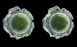 Creative Commons Attribution License; PLoS ONE 2010;5:e10659
This stereo image of a lens from a 64-year-old male subject with Down syndrome shows a characteristic circumferential supranuclear cataract presenting as an annular half-toroid band of opacification in the deep cortical and supranuclear subregions of the lens.
"The mouse had a cataract. I looked at the other eye, and there was a cataract there, too. That’s very unusual – really not ever seen – in a mouse this age."
Then he looked at all the other mice he was using in that experiment, all of which were older. They all had bilateral cataracts. My first thought was, "This can’t be related to Alzheimer’s disease."
But in fact, he said, it appeared to be. He and his lab soon showed that the cataracts contained a large concentration of aggregated beta-amyloid (Abeta) in the same fraction that’s measured in today’s cerebrospinal fluid Alzheimer’s biomarker tests.
That first observation has birthed two investigational noninvasive amyloid eye tests, which Dr. Goldstein envisions could some day be part of everyone’s annual physical exam.
In people destined to develop Alzheimer’s, some research suggests that Abeta proteins may begin to accumulate in the lens long before they build up to dangerous levels in the brain. If this turns out to be a reliable marker of risk, it could be a sign that would trigger early, presymptomatic Alzheimer’s treatment.
That’s in the future, though, because right now, there is no such treatment. But at this time, Dr. Goldstein said, an amyloid eye test could prove invaluable in reaching that goal. One reason that symptomatic patients don’t respond to investigational drugs could be that by the time the patients are treated, irreversible brain damage has already occurred.
"Once you have cognitive symptoms, the horse is not only out of the barn, it’s run out of the state," said Dr. Goldstein, director of the molecular aging and development laboratory at Boston University. "I hate the term ‘mild cognitive impairment,’ because by the time you have that, there’s nothing mild about it."
Researchers now almost universally agree that the best way to get a true picture of any drug’s potential effectiveness in Alzheimer’s will be to implement treatment before symptoms set in. In addition, Dr. Goldstein said, "Research pools are polluted. Control groups contain subjects who would develop Alzheimer’s if they live long enough," which could be skewing study results. Lens amyloid measurements might help stratify groups in drug studies, and even be a way to track very early effect on amyloid.
But that is a future yet to be determined. In the meantime, researchers still need definitive proof that supranuclear amyloid cataracts are inextricably linked to the amyloid brain plaques of Alzheimer’s.
Initial findings
In 2003, Dr. Goldstein, then at Harvard Medical School, published his original proof of concept study. It comprised postmortem eye and brain specimens from nine subjects with Alzheimer’s and eight controls without the disease, and samples of primary aqueous humor from three people without the disorder who were undergoing cataract surgery (Lancet 2003;361:1258-65).
Abeta-40 and Abeta-42 were present in all of the lenses, in amounts similar to those seen in the corresponding brains. But in patients with Alzheimer’s, the protein aggregated into clumps within the lens fiber cells, forming unusual supranuclear cataracts at the equatorial periphery that appeared to be different from common age-related cataracts and that weren’t present in the control subjects.
The cataract location is an important clue to how long the Abeta has been accumulating, Dr. Goldstein said. Lens fiber cells are particularly long lived, remaining alive for as long as a person lives or until the lens is removed during cataract surgery. The lens starts to form in very early fetal life, with more and more lens cells forming in an outward direction, creating a virtual map of a person’s lifetime, "like the rings of a tree," he said.
Dr. Goldstein and his team discovered that these distinctive cataracts develop in some patients with Alzheimer’s. They appear toward the outer edge of the lens and are composed of the same toxic Abeta protein that builds up in the brain. "The history of amyloid in the body is time stamped in the lens," he said.