Conference Coverage


FROM ANNALS OF THE RHEUMATIC DISEASES
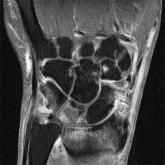
The joints of patients with early arthritis that showed subclinical inflammation on MRI had an increased risk of radiographic progression in the first year after disease presentation, particularly those with bone marrow edema, according to findings from the first study to look at the radiographic outcome of such joints.
A total of 179 patients at the Leiden Early Arthritis Clinic, a population-based inception cohort including patients with confirmed clinical arthritis and symptoms for less than 2 years, underwent 1.5T extremity MRI at baseline of the metacarpophalangeal 2-5, wrist, and metatarsophalangeal 1-5 joints on the side with the most severe symptoms or the dominant side in cases in which both sides were equally affected. At 1-year follow-up, 113 had radiographs taken. Close to half of these patients (46.9%) fulfilled the 2010 ACR/EULAR criteria for rheumatoid arthritis (RA) at baseline, and those who did not have follow-up at 1 year were diagnosed with RA less often. Other patients had unclassified arthritis (32.7%), psoriatic arthritis (9.7%), inflammatory osteoarthritis (3.5%), spondylarthritis (2.7%), or other diagnoses (4.4%). Within the first year, 77% of all patients received treatment with a disease-modifying antirheumatic drug, including 93% of all patients with RA, said Dr. Annemarie Krabben and her colleagues at Leiden University Medical Center, the Netherlands (Ann. Rheum. Dis. 2014 July 29 [doi:10.1136/annrheumdis-2014-205208]).
Of the 1,130 joints studied, 932 were clinically free of swelling at baseline, and, of these, 232 (26%) had subclinical inflammation on MRI, including 17% with bone marrow edema, 16% with synovitis, and 21% with tenosynovitis. Overall, only 2% of the unswollen joints had radiographic progression at 1 year, compared with 8% of clinically swollen joints. The relative risk for radiographic progression in unswollen joints with subclinical inflammation on MRI was 3.5 (95% confidence interval, 1.3-9.6) based on radiographic progression in 4% of those with subclinical inflammation and 1% of those without it. The relative risk was highest for nonswollen joints with bone marrow edema (RR, 5.3; 95% CI, 2.0-14.0) and also significant for those with synovitis (RR, 3.4; 95% CI, 1.2-9.3), but not for those with tenosynovitis (RR, 3.0; 95% CI, 0.7-12.7). The relative risk was comparable for joints that were both nonswollen and nontender, although the 95% CI was broader.
"Our observation that the total level of MRI inflammation [in both swollen and unswollen joints] was an independent predictor for radiographic progression fits with previous findings," the investigators wrote. "Furthermore, our finding that subclinical inflammation at disease onset is associated with radiographic progression is in line with previous findings on subclinical inflammation in patients with RA in clinical remission."
The authors noted that their "study mainly increases the comprehension of the connection between inflammation and structural damage early in the disease. Whether subclinical MRI inflammation is relevant to clinical practice remains a question, as rheumatologists treat patients and not joints. Information on subclinical inflamed joints would affect treatment decisions most when patients have few clinically swollen joints."
The size of the study’s patient sample limits the applicability of the findings, particularly in regard to the moderate number of patients with RA, who had broad 95% CI estimates for the risk of radiographic progression. Other limits to the study included the relative infrequency of radiographic progression at 3% of all joints (4% of joints in patients with RA) and the relatively small increase of 1 or greater on the Sharp-van der Heijde score that was used to define radiographic progression.
The study was supported by the Dutch Arthritis Foundation, the Dutch Organization of Health Research and Development, the Center for Translational Molecular Medicine, and by a grant within the European Community Seventh Framework Programme FP7 (Masterswitch). The authors had no competing interests to declare.

