Gynecologic Oncology Consult

Approaching intraoperative bowel injury


Enterotomy can be a serious complication in abdominopelvic surgery, particularly if it is not immediately recognized and treated. Risk of visceral injury increases when complex dissection is required for treatment of cancer, resection of endometriosis, and extensive lysis of adhesions.

In a retrospective review from 1984 to 2003, investigators assessed intestinal injuries at the time of gynecologic operations. Of the 110 cases reported, about 37% occurred during the opening of the peritoneal cavity, 38% during adhesiolysis and pelvic dissection, 9% during laparoscopy, 9% during vaginal surgery, and 8% during dilation and curettage. Of the bowel injuries, more than 75% were minor.1 Mortality from unrecognized bowel injury is significant, and as such, appropriate recognition and management of these injuries is critical.2

Dr. Allison Staley of University of North Carolina, Chapel Hill.

Dr. Allison Staley

The wall of the small intestine, from in to out, consists of layers: the mucosa, muscularis, and serosa. The muscularis layer is composed of an inner circular muscle and outer longitudinal muscle. The posterior parietal peritoneum encloses the bowel to form the mesentery and provide covering for the vasculature, lymphatics, and nerves supplying the small intestine. The arterial supply for the jejunum and ileum originates from the superior mesenteric artery. Branches within the mesentery anastomose to form arcades. The straight arteries from these arcades supply the mesenteric border of the gut.3 Familiarity with bowel anatomy is important in order to accurately diagnose the extent of injury and determine the optimal repair technique.

Some basic principles are critical when surgeons face a bowel injury:

1. Recognize the extent of the injury, including the size of the breach, the depth (full or partial thickness), and the nature of the injury (thermal or cold).

2. Assess the integrity of the bowel, including adequacy of blood supply, prior bowel damage from radiation, and absence of downstream obstruction.

3. Ensure no other occult injuries exist in other segments.

4. Obtain adequate exposure and mobilization of the bowel beyond the site of injury, including the adjacent bowel. This involves releasing other adhesions so that adequate bowel length is available for a tension-free repair.

Methods of repair

The two main methods of bowel repair are primary closure and resection with re-anastamosis. The decision to employ each is influenced by multiple factors. Primary closure is best suited to small lesions (1 cm or less) that are a result of cold or sharp injury. However, thermal injury sustained via electrosurgical devices induces delayed tissue damage beyond the visible edges of the immediate defect, and surgeons should consider a resection of bowel to at least 1 cm beyond the immediately apparent injury site. Additionally, resection and re-anastamosis should also be considered if the damaged segment of bowel has poor blood supply, integrity, or the repair would result in tension along the suture/staple line or luminal narrowing.

Simple small bowel closures

Serosal abrasions need not be repaired; however, small tears of the serosa and muscularis can be managed with a single layer of interrupted 3-0 absorbable or permanent silk suture on a tapered needle. The suture line should be perpendicular to the longitudinal axis of the bowel at 2-mm to 3-mm intervals in order to prevent narrowing of the lumen. The suture should pass through serosal and muscular layers in an imbricating (Lembert) stitch. For smaller defects of less than 6 mm, a single layer closure is typically adequate.

The suture line should be perpendicular to the longitudinal axis of the bowel and the suture should pass through serosal and muscular layers in Lembert stitches.

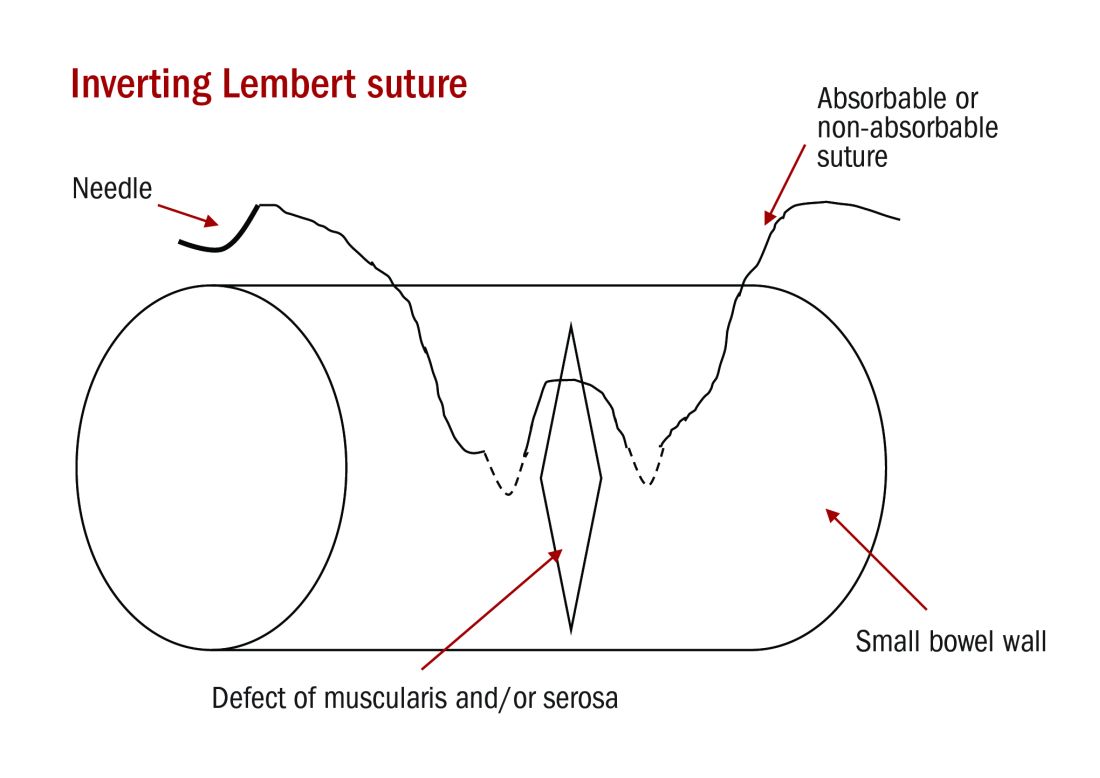
Small tears can be repaired with a single layer of interrupted 3-0 absorbable or permanent silk suture on a tapered needle.

For full thickness and larger single defects, a double layer closure is recommended with a full-thickness inner layer (including the mucosa) in which the mucosa is inverted luminally with 3-0 absorbable suture in a running or interrupted fashion followed by a seromuscular outer layer of 3-0 absorbable or silk sutures placed in interrupted imbricating Lembert stitches. Care should be taken to avoid stricture of the lumen and tearing of the fragile serosal tissue. Sutures placed in an interrupted fashion as opposed to continuous or “running” sutures are preferred because they reapproximate tissues with less tissue necrosis and less chance for luminal narrowing. Antibiotics need not be prescribed intraoperatively for a small bowel breach.

Small bowel resection

Some larger defects, thermal injuries, and segments with multiple enterotomies may be best repaired with resection and re-anastamosis technique. A segment of resectable bowel is chosen such that the afferent and efferent limbs to be re-anastamosed can be reapproximated in a tension-free fashion. A mesenterotomy is made at the proximal and distal portions of the involved bowel. A gastrointestinal anastomotic stapler is then inserted perpendicularly across the bowel. The remaining wedge of connected mesentery can then be efficiently excised with an electrothermal bipolar coagulator device ensuring that maximal mesentery and blood supply are preserved to the remaining limbs of intestine. The proximal and distal segments are then aligned at the antimesenteric sides.

Dr. Emma C. Rossi is an assistant professor in the division of gynecologic oncology at UNC-Chapel Hill.

Dr. Emma C. Rossi

To assist with stabilization, a simple silk suture may be placed through the antimesenteric border of the segments. The corner of each segment on the antimesenteric side is incised just enough to cut through all three layers of the bowel wall. Each GIA stapler limb is passed through the proximal and distal segments. These are then aligned on the antimesenteric sides and the GIA stapler is closed and deployed. The final step is closure of the remaining enterotomy. This is grasped with Allis clamps, and a line of staples – typically either a transverse anastomosis stapler or another application of the GIA stapler – is placed around the bowel just beneath the Allis clamps and excess tissue is sharply trimmed. The mesenteric defect must also be closed prior to completion of the procedure to avoid internal herniation of the bowel or omentum. This may be closed with running or interrupted delayed-absorbable suture.4,5

Pages

Recommended Reading

Understanding the human papillomavirus
MDedge ObGyn
Use of Pap smears, mammography on the decline
MDedge ObGyn
Optimizing HPV vaccination
MDedge ObGyn
Low malignant potential tumors of the ovary: A review
MDedge ObGyn
Does HPV testing lead to improved diagnosis of cervical dysplasia for patients with ASC-US cytology?
MDedge ObGyn
Is pain or dependency driving elevated opioid use among long-term cancer survivors?
MDedge ObGyn
FDA approves biosimilar to bevacizumab
MDedge ObGyn
HPV vaccine pioneers win 2017 Lasker-DeBakey Clinical Medical Research Award
MDedge ObGyn
Surviving ovarian cancer: Is there an association between hospital volume and quality of care?
MDedge ObGyn
Breast density and optimal screening for breast cancer
MDedge ObGyn