Multiple gestations are far more common today than they once were—up 70% since 1980.1 Today, every 1,000 live births include 32.3 sets of twins, a 2% increase in the rate of twin births since 2004. Why this phenomenon is occurring is not entirely understood but, certainly, the trend toward older maternal age and the emergence of assisted reproduction are both part of the explanation.
Multiple gestations are of particular concern to obstetricians because, even though they remain relatively rare, they are responsible for a significant percentage of perinatal morbidity and mortality.
The difficulties that twins encounter are often associated with preterm birth and occur most often in identical twins developing within a single gestational sac. Those difficulties include malformation, chromosomal abnormalities, learning disability, behavioral problems, chronic lung disease, neuromuscular developmental delay, cerebral palsy, and stillbirth. Women pregnant with twins are also at heightened risk, particularly of gestational hypertension, preeclampsia, and gestational diabetes.2
Your task is to manage these risks so that the outlook for mother and infant is as favorable as possible.
Determining chorionicity in the first trimester
Ultrasonographic determination of chorionicity should be the first step in the management of a twin gestation. The determination should be made as early as possible in the pregnancy because it has an immediate impact on counseling, risk of miscarriage, and efficacy of noninvasive screening. (See “Is there 1 sac, or more? Key to predicting risk.”)
The accuracy of ultrasonography (US) in determining chorionicity depends on gestational age. US predictors of dichorionicity include:
- gender discordance
- separate placentas
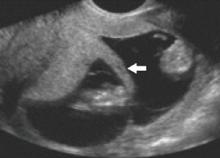
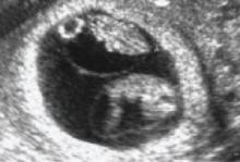
- the so-called twin-peak sign (also called the lambda sign) (FIGURE 1), in which the placenta appears to extend a short distance between the gestational sacs; compare this with FIGURE 2, showing monochorionic twins with the absence of an intervening placenta
- an intertwin membrane thicker than 1.5 mm to 2.0 mm.
US examination can accurately identify chorionicity at 10 to 14 weeks’ gestation, with overall sensitivity that is reported to be as high as 100%.3-5
FIGURE 1
The twin-peak sign on US
This dichorionic–diamnionic twin gestation demonstrates the so-called twin peak, or lambda, sign (arrow), in which the placenta appears to extend a short distance between the gestational sacs.
FIGURE 2
Monochorionic–diamnionic twin gestation
This US scan of a monochorionic twin gestation reveals the absence of an intervening placenta
Monochorionic twins
Twins who share a gestational sac are more likely than 2-sac twins to suffer spontaneous loss, congenital anomalies, growth restriction and discordancy, preterm delivery, and neurologic morbidity.
Spontaneous loss. In 1 comparative series, the risk of pregnancy loss at less than 24 weeks’ gestation was 12.2% for monochorionic twins, compared with 1.8% for dichorionic twins.6 Spontaneously conceived monochorionic twins may have the highest risk of loss.7 However, monochorionic twins occur more often in conceptions achieved by assisted reproductive technology—at a rate 3 to 10 times higher than the background rate of monochorionic twinning.8
- A multiple gestation involves a higher level of risk than a singleton pregnancy
- Chorionicity is the basis for determining risk. Twins within a single sac (monochorionic) are at higher risk of malformation, Down syndrome, and premature birth
- The risk of Down syndrome can be estimated by noninvasive screening in the first trimester and by chorionic villus sampling or amniocentesis later in the pregnancy
- A detailed anatomic survey at 18 to 20 weeks’ gestation should be done to detect possible malformations
- Assessment of cervical length, performed every 2 weeks from the 16th to the 28th week, may help predict premature delivery—but is not definitive
- Assessment of fetal growth every 4 weeks in dichorionic twins and every 2 weeks in monochorionic twins can alert you to potential problems. This is particularly important for detecting signs of twin-to-twin transfusion syndrome
Congenital anomalies. These occur 2 to 3 times as often in monochorionic twins, and have been reported in as many as 10% of such pregnancies. Reported anomalies include midline defects, cloacal abnormalities, neural tube defects, ventral wall defects, craniofacial abnormalities, conjoined twins, and acardiac twins.9-11 In light of these risks, a detailed anatomic survey is suggested for all twins.


