Surgical Techniques

Step by step: Obliterating the vaginal canal to correct pelvic organ prolapse

Author and Disclosure Information

Very elderly age, comorbidity, and disinterest in maintaining sexual function make a woman an ideal candidate for having POP corrected by surgery to close the vaginal canal, detailed here


This article, with accompanying video footage, is presented with the support of the International Academy of Pelvic Surgery.

As women live longer, on average, pelvic floor disorders are, as a whole, becoming more prevalent and a greater health and social problem. Many women entering the eighth and ninth decades of life display symptomatic pelvic organ prolapse (POP)—often after an unsuccessful trial of a pessary or even surgery.

These elderly patients often have other concomitant medical issues and are not sexually active, making extensive surgery for them less than an ideal solution. Instead, surgical procedures that obliterate the vaginal canal can alleviate their symptoms of POP.

In this article, we provide a step-by-step description of:

  • LeFort partial colpocleisis in a woman who still has her uterus in place
  • partial or complete colpectomy and colpocleisis in a woman who has post-hysterectomy prolapse
  • levator plication and perineorrhaphy, as essential concluding steps in these procedures.

LeFort partial colpocleisis

An obliterative procedure in the form of a LeFort partial colpocleisis is an option when a patient 1) has her uterus and 2) is no longer sexually active. Because the uterus is retained in this procedure, however, keep in mind that it will be difficult to evaluate any uterine bleeding or cervical pathology in the future. Endovaginal ultrasonography or an endometrial biopsy, and a Pap smear, must be done before LeFort surgery.

The ideal candidate for LeFort partial colpocleisis is a woman who has complete uterine prolapse, or procidentia (FIGURE 1), which is characterized by symmetric eversion of the anterior and posterior vaginal walls.


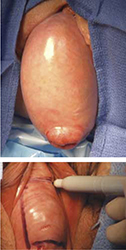
FIGURE 1 Pelvic organ prolapse, preoperatively
Top: Uterine procidentia. A patient who has this condition is an ideal candidate for LeFort partial colpocleisis. Bottom: Asymmetric anterior vaginal prolapse.

LeFort partial colpocleisis: Key step by key step

Begin by placing the cervix on traction to evert the vagina. Inject the vaginal mucosa with either bupivacaine or 2% lidocaine with 1:200,000 epinephrine, just below the vaginal epithelium. Place a Foley catheter with a 5-mm balloon into the bladder so that you can identify the bladder neck.
Use a marking pen to mark out the rectangular areas of the vaginal epithelium that are to be removed anteriorly and posteriorly. Extend the anterior rectangle from approximately 2 cm from the tip of the cervix to 4 or 5 cm below the external urethral meatus. Mark out a mirror image on the posterior aspect of the cervix and vagina. Extend the rectangle on the posterior vaginal wall from approximately 2 cm below the level of the tip f the cervix to 4 or 5 cm inside the posterior fourchette.
Incise the previously marked areas and utilize sharp dissection to remove the vaginal epithelium from both the anterior and posterior vaginal walls. Leave the maximum amount possible of vaginal muscularis on the underlying bladder and the rectum. Hemostasis is an absolute must. When you remove the posterior vaginal flap, avoid entering the peritoneum; if you do enter it inadvertently, close the defect with interrupted delayed absorbable suture.
Sew together the cut edges of the anterior and posterior vaginal walls with interrupted delayed absorbable sutures. When possible, turn the knot into the epithelium-lined tunnels that you have created bilaterally. Turn the uterus and vaginal apex gradually inward. After the vagina has been inverted, suture the superior and inferior margins of the rectangle together.
Our opinion is that a support procedure—at either the bladder neck (Kelly plication) or midurethra (synthetic midurethral sling)—should be performed on all patients, based on preoperative assessment for potential or occult urinary stress incontinence. For more discussion, see Question 7 in “Questions we’re asked (and answers we give) about obliterative surgery.”
Perform levator plication and perineorrhaphy as a matter of routine. Key steps in these procedures are provided in the final section of the article.
Postoperatively, the patient is mobilized early, although she should avoid heavy lifting for at least 6 weeks to prevent recurrence of the prolapse secondary to breakdown of the repair.

FIGURE 2 shows key steps in performing LeFort partial colpocleisis. See Video #1 at www.obgmanagement.com for demonstrations of how to perform LeFort partial colpocleisis.


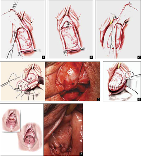
FIGURE 2 Steps: LeFort partial colpocleisis
A. Denude the anterior vaginal epithelium. B. Plicate the neck of the bladder. C. Next, denude the posterior vaginal epithelium. D. Approximate most proximal surfaces. E. Place lateral sutures to allow for drainage canals. F. The uterus has been replaced and most of the distal incisions closed.

Pages

Recommended Reading

Robotic Hysterectomy Found Safe in Morbidly Obese
MDedge ObGyn
Early Oophorectomy Linked to Osteoporosis, Arthritis
MDedge ObGyn
Early Bilateral Oophorectomy Is Linked to Osteoporosis, Arthritis
MDedge ObGyn
Robotic Hysterectomy Safe in Morbidly Obese
MDedge ObGyn
Longer OR Times Confirmed in Robotic Hysterectomy
MDedge ObGyn
Outpatient Hysterectomy Results Prove Positive
MDedge ObGyn
How to repair bladder injury at the time of cesarean delivery
MDedge ObGyn
What is the recommended approach to a breast mass in a woman younger than 25 years?
MDedge ObGyn
UPDATE ON URINARY INCONTINENCE
MDedge ObGyn
Is energy-based vessel sealing safer than suturing for vaginal hysterectomy?
MDedge ObGyn