Physicians and patients heading into the operating room have to hope that the surgeon or proceduralist does the job right the first time. Generally, needing a do-over is not a good thing.
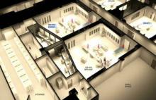
Soon, however, vascular and cardiac surgeons at Cleveland Clinic will be able to take several consequences-free do-overs that should lower risk for the patient. Some of the latest advances in imaging and technology are being incorporated into new simulation rooms built adjacent to a few new ORs. In these rooms physicians can rehearse the procedure they’re about to do on a three-dimensional simulation of the patient, who is being prepared for surgery next door.
For example, “fusion” imaging can help in the repair of aortic aneurysms. The results of an angiogram taken at the time of the procedure are superimposed on a preoperative CT scan of the patient to create three-dimensional imaging studies “telling us where all the arteries are without using any contrast,” Dr. Roy K. Greenberg said at the annual meeting of the Southern Association for Vascular Surgery. Not having to use catheters and wires to figure that out makes the procedure much easier, said Dr. Greenberg of the Cleveland Clinic Foundation.
His talk on “Aortic Care for the 21st Century” at the meeting was the first Jesse E. Thompson, M.D. Distinguished Guest Lecture.
“The imaging can be loaded in patient-specific simulators, providing a means to ‘test’ the procedures in virtual reality, train the team, and problem-solve `off-line’ ” with less risk to the patient, he said.
The Cleveland Clinic is no slouch when it comes to simulation. Their doctors-in-training practice on mannequins, robots, and other virtual stand-ins for patients. There is an Anesthesia Simulation Lab. The Clinic is even building an entire Center for Multidisciplinary Simulation.
The two new ORs that Dr. Greenberg described take simulation to the next level. The simulators are not in a remote room, they’re not in a separate building, and they’re not at a course that physicians go to for two or three days before the surgery. “I think they have to be integrated into the actual work flow and case,” he said.
The ORs should be finished in the fourth quarter of 2012, and will feature a control room behind each OR. Behind each control room will be a simulator room.
Dr. Greenberg described the work flow for a fenestrated endovascular aneurysm repair as something like this: The preoperative CT scan is used for patient selection and device design, and is imported into the OR and the simulator before the patient arrives. The CT scan and fluoroscopic imaging studies are used in fusion imaging in the OR, minimizing the use of contrast agents and radiation.
Standard clinic procedures required team sign-ins when the patient is on the table. Each team members signs in, introduces themselves, and makes sure they're operating on the correct anatomic part. Then “we have that obligatory 2.5 hours while anesthesia gets everything else ready,” during which the operative team goes to the simulator room. Residents, fellows, and the rest of the team get to practice the procedure using the repair device on a simulation of that particular patient.
“So there’s no question about what steps are next” during the live surgery, he said. If something doesn’t seem right during rehearsal, “you can say, `Why not?’ Is that a problem with the simulator, or is it going to be a problem with the procedure?”
Dr. Greenberg said he doesn’t know of any other institutions doing patient-specific simulations like this, but “I hope they will.”
Dr. Greenberg disclosed receiving royalties for intellectual property licensed to Cook, Inc. and funds for research and travel from Cook.

