SKIN MANIFESTATIONS ASSOCIATED WITH TYPE 2 DIABETES
Yellow nails
Elderly type 2 diabetic patients tend to have yellow nails. A prevalence of 40% to 50% in patients with type 2 diabetes has been reported,14 but occasionally yellow nails are also found in normal elderly people and in patients with onychomycosis. The yellow discoloration in diabetes is most evident on the distal end of the hallux nail. It probably represents end-products of glycosylation, similar to the yellow color in diabetic skin, although this has not yet been confirmed.15
Diabetic thick skin
Diabetes mellitus is generally associated with a thickening of the skin,2 measurable via ultrasonography,16 and this thickening may increase with age in all diabetic patients, unlike normally aging skin.
Diabetic thick skin occurs in three forms. First is the general asymptomatic but measurable thickening. Second is a clinically apparent thickening of the skin involving the fingers and hands. Third is diabetic scleredema, an infrequent syndrome in which the dermis of the upper back becomes markedly thickened.2,6
Thickening of the skin on the dorsum of the hands occurs in 20% to 30% of all diabetic patients, regardless of the type of diabetes.17 Manifestations range from pebbled knuckles to diabetic hand syndrome.2 Pebbled knuckles (or Huntley papules) are multiple minute papules, grouped on the extensor side of the fingers, on the knuckles, or on the periungual surface.18 The prevalence of diabetic hand syndrome varies from 8% to 50%.19 It begins with stiffness of the metacarpophalangeal and proximal interphalangeal joints and progresses to limit joint mobility.20,21 Dupuytren contracture (or palmar fascial thickening) may further complicate diabetic hand syndrome.5,22
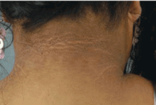
Scleredema diabeticorum is characterized by remarkable thickening of the skin of the posterior neck and upper back, occasionally extending to the deltoid and lumbar regions. A peau d’orange appearance of the skin can occur, often with decreased sensitivity to pain and touch.
Scleredema occurs in 2.5% to 14% of people with diabetes6 and is sometimes confused with scleredema of Buschke, a rare disorder in which areas of dermal thickening occur, mostly on the face, arms, and hands, often after an upper respiratory infection. It clears spontaneously in months or years. Women are affected more often than men. These characteristics differentiate scleredema of Buschke from scleredema diabeticorum, which almost exclusively occurs in long-standing diabetes, is usually permanent, is not related to previous infection, and is limited to the posterior neck and upper back. No effective treatment is known for scleredema diabeticorum.23
Skin tags or acrochordons
Skin tags are small, pedunculated, soft, often pigmented lesions occurring on the eyelids, the neck, and the axillae. A few studies have reported an association between multiple skin tags and diabetes, and between skin tags and insulin resistance.24–27 Crook28 found that skin tags were associated with the typical atherogenic lipid profile seen in insulin-resistant states: elevated triglycerides and low levels of high-density-lipoprotein cholesterol. In a large study of patients with skin tags,24 over 25% had diabetes and 8% had impaired glucose tolerance.24
Treatment is not necessary, but skin tags can be removed with grade 1 scissors, cryotherapy, or electrodessication.28 Skin tags may be regarded as a sign of impaired glucose tolerance, diabetes, and increased cardiovascular risk.28,29
Diabetic dermopathy
Diabetic dermopathy (ie, shin spots and pigmented pretibial papules) affects 7% to 70% of all diabetic patients. It is not specific for diabetes: 20% of nondiabetic people show similar lesions. Men are affected more often than women, and the mean age is 50 years.
Shin spots present as multiple, bilateral, asymmetrical, annular or irregular red papules or plaques on the extensor surface of the lower legs and may precede abnormal glucose metabolism. The clinician usually sees only the end result: atrophic, scarred, hyperpigmented, finely scaled macules. Lesions may also be found on the forearms, thighs, and lateral malleoli. Several studies found severe microvascular complications in patients with diabetic dermopathy, indicating a close association with a high risk of accelerated diabetes complications.
Treatment is not very effective; however, some lesions resolve spontaneously.6,30
Acanthosis nigricans
The pathogenesis is most likely related to high levels of circulating insulin, which binds to insulin-like growth factor receptors to stimulate the growth of keratinocytes and dermal fibroblasts.
Although the lesions are generally asymptomatic, they can be painful, malodorous, or macerated.3 The most effective treatment is lifestyle alteration. Weight reduction and exercise can reduce insulin resistance. Acanthosis nigricans is reversible with weight reduction if it is seen as a complication of obesity. If the lesions are asymptomatic, they need no treatment. Ointments containing salicylic or retinoic acid can be used to reduce thicker lesions in areas of maceration in order to decrease odor and promote comfort. Systemic isotretinoin (Accutane) improves acanthosis nigricans, but it recurs when the drug is discontinued.3,5,6,32
Acquired perforating dermatosis
Acquired perforating dermatosis is seen in patients with kidney failure, type 2 diabetes, or type 1 diabetes. A prevalence of up to 10% has been reported in dialysis patients.33,34
The characteristic lesions are 2- to 10-mm, pruritic, dome-shaped papules and nodules with a hyperkeratotic plug. They occur mainly on the limbs, the trunk, and the dorsal surface of the hands, and to a lesser extent on the face. The Koebner phenomenon (also called isomorphic effect) may also occur.
Histologic study shows a hyperplastic epidermis with marked spongiosis directly over the plug. The contents of the plug itself are collagen, elastic fibers, nuclear debris, and polymorphonuclear leukocytes. These leukocytes have been implicated in the pathogenesis of acquired perforating dermatosis.6,17
The lesions are chronic but may heal after months if trauma and scratching are avoided. Further treatments include topical keratolytics, psoralen-ultraviolet A light, ultraviolet B light, topical and systemic retinoids, topical and intralesional steroids, oral antihistamines, and cryotherapy.6


