Case Reports

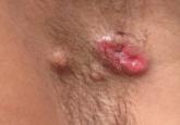
Primary Apocrine Adenocarcinoma of the Axilla
Primary apocrine adenocarcinoma (AA) is a rare malignant cutaneous neoplasm that typically arises in areas of high apocrine gland density such as...
Chayada Kokpol, MD; Emily Y. Chu, MD, PhD; Suthinee Rutnin, MD
Drs. Kokpol and Rutnin are from the Division of Dermatology, Ramathibodi Hospital, Mahidol University, Bangkok, Thailand. Dr. Chu is from the Department of Dermatology, Hospital of the University of Pennsylvania, Philadelphia.
The authors report no conflict of interest.
Correspondence: Suthinee Rutnin, MD, Division of Dermatology, Department of Medicine, Ramathibodi Hospital, Mahidol University, 270 Rama VI Rd, Ratchatewi, Bangkok, Thailand 10400 (kungkling_107@yahoo.com).

Nodular scleroderma is a rare form of scleroderma that may occur systemically or locally. The pathogenesis of this variant is unknown. We report the case of a 63-year-old woman with systemic scleroderma and chronic hepatitis C virus (HCV) infection who had numerous papules and nodules on the neck and trunk. Skin biopsies from her lesions revealed characteristic findings of scleroderma. This case not only depicts the rare entity of nodular scleroderma but demonstrates the association of HCV infection with systemic autoimmune diseases such as scleroderma.
Practice Points
Case Report
A 63-year-old woman was referred to our clinic for evaluation of multiple papules and nodules on the neck and trunk that had been present for 2 years. Three years prior to presentation she had been diagnosed with systemic sclerosis (SSc) after developing progressive diffuse cutaneous sclerosis, Raynaud phenomenon with digital pitted scarring, esophageal dysmotility, myositis, pericardial effusion, and interstitial lung disease. Serologic test results were positive for anti-Scl-70 antibodies. Antinuclear antibody test results were negative for anti–double-stranded DNA, anti-nRNP, anti-Ro/La, anti-Sm, and anti-Jo-1 antibodies. The patient was treated with prednisolone 7.5 mg daily, nifedipine 15 mg daily, valsartan 80 mg daily, manidipine 20 mg daily, omeprazole 20 mg daily, and beraprost 80 mg daily. One year later, numerous asymptomatic flesh-colored papules and nodules developed on the neck, chest, abdomen, and back. There was no history of trauma or surgery at any of the affected sites.
On further investigation, anti–hepatitis C virus (HCV) antibodies were identified and confirmed by HCV ribonucleic acid polymerase chain reaction at the same time that the diagnosis of SSc was established. Hepatitis C virus genotype 3a was noted, and the patient’s viral load was 378,000 IU/mL. Therefore, a diagnosis of chronic HCV infection was established. The patient was initially unable to receive medical treatment due to lack of finances. A year and a half following the diagnosis of HCV infection, with worsening liver function tests and increasing viral load (1,369,113 IU/mL), the patient began therapy with peginterferon alfa-2b 80 mg weekly and ribavirin 800 mg daily. However, the medications were discontinued after 2 months when she developed severe hemolytic anemia related to ribavirin.
On physical examination, the patient was noted to have a masklike facies with a pinched nose and constricted opening of the mouth. Her skin was tightened and stiff extending from the fingers to the proximal extremities. Numerous well-circumscribed, flesh-colored, firm papules and nodules ranging from 2 to 20 mm in diameter were present on the neck (Figure 1), chest, abdomen (Figure 2), and back.
| Figure 1. Numerous flesh-colored firm papules and nodules on the posterior aspect of the neck. | Figure 2. Multiple well-defined sclerotic papules and nodules on the abdomen. |
Two 4-mm punch biopsy samples obtained from a papule on the neck and a nodule on the abdomen revealed homogenized collagen bundles with scattered plump fibroblasts in the lower reticular dermis. Clinicopathologic correlation of the biopsy findings with the cutaneous examination resulted in a diagnosis of nodular scleroderma (Figures 3 and 4).
Figure 3. The collagen bundles in the reticular dermis appeared thickened and closely packed. They stained more deeply eosinophilic than in the upper dermis. The overlying epidermis was normal (H&E, original magnification ×4). Figure 4. Thick, pale, hyalinized collagen bundles with scattered fibroblasts were seen in the lower reticular dermis. The eccrine glands were surrounded by sclerotic collagen and only a few adipocytes (H&E, original magnification ×20). |
The patient began treatment with intralesional injections of triamcinolone 5 to 10 mg/mL for nodules as well as an ultrapotent corticosteroid cream, clobetasol propionate 0.05%, for small papules. Injections were performed at 4- to 8-week intervals and resulted in modest clinical improvement.
Comment
Scleroderma may be present only in the skin (morphea) or as a systemic disease (systemic scleroderma). Rarely, cutaneous involvement can exhibit a nodular or hypertrophic morphology, which has been described in the literature as nodular or keloidal scleroderma in a patient with known SSc1-10 and as nodular or keloidal morphea in localized cutaneous scleroderma.3,11-13
Histopathology
The distinction between the terms nodular scleroderma and keloidal scleroderma is not clear, and they are not necessarily interchangeable. To provide clarity, we find it useful to delineate specific histologic findings associated with the diagnoses of keloid, scleroderma, and the uncommon keloid/scleroderma overlap. The histopathologic findings of keloids include a fibrotic dermis and broad dispersed bundles of eosinophilic hyalinized collagen. The histopathologic findings of scleroderma include broad sclerotic bands of collagen throughout the dermis with loss of perieccrine fat. In the overlapping keloid/scleroderma condition, which is a variant of scleroderma, hyalinized collagen fibers and keloidal collagen appear in the same specimen.3,4
To distinguish these conditions, Barzilai et al5 proposed that only cases showing both clinical and histologic characteristics of a keloid should be referred to as keloidal morphea/scleroderma. They further stated that the terms nodular morphea or nodular scleroderma ought to be used only for cases that are indistinguishable histologically from scleroderma. The term morphea is appropriate when only a limited amount of skin disease is present, while scleroderma implies association with systemic disease.5 Likely, there is a histologic continuum in this variant of scleroderma, in which nodular morphea/scleroderma exists at one end and keloidal morphea/scleroderma exists at the other end.5,13

Primary apocrine adenocarcinoma (AA) is a rare malignant cutaneous neoplasm that typically arises in areas of high apocrine gland density such as...
The ulcerative variant of lichen planus (LP) commonly involves the oral mucosa but is uncommon and difficult to treat when located on other areas...
Various factors contribute to the development of porphyria cutanea tarda (PCT). In this case report, we describe a patient with hepatitis C...
