All from the University of Mississippi Medical Center, Jackson. Dr. Brodell also is from the School of Medicine and Dentistry, University of Rochester Medical Center, New York.
The authors report no conflict of interest.
Correspondence: Robert T. Brodell, MD, Division of Dermatology, University of Mississippi Medical Center, 2500 N State St, Jackson, MS 39216 (rbrodell@umc.edu).
Porokeratosis of Mibelli (PM) is a rare condition with the potential for malignant transformation that presents a clinical and pathologic diagnostic challenge. An improperly oriented biopsy may lead to the wrong histopathologic diagnosis. We report a case of PM that was previously misdiagnosed and describe a biopsy technique for suspected PM that maximizes the potential for histopathologic confirmation of the diagnosis.
A biopsy from the center of a plaque of porokeratosis will produce nonspecific findings.
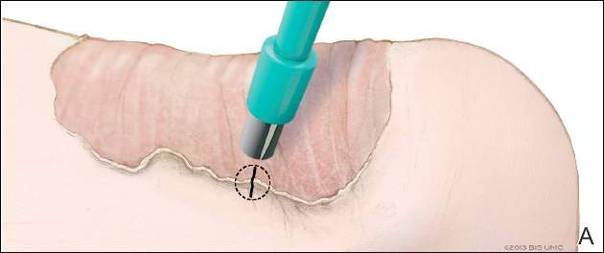
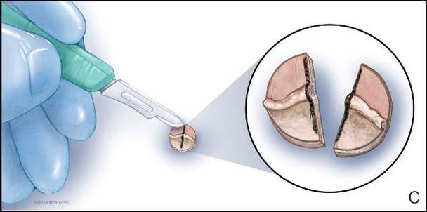
Bisecting the punch specimen at the bedside along a line drawn perpendicular to the cornoid lamella guarantees proper orientation of the specimen.
References
Porokeratosis of Mibelli (PM) is a lesion characterized by a surrounding cornoid lamella with variable nonspecific findings (eg, atrophy, acanthosis, verrucous hyperplasia) in the center of the lesion that typically presents in infancy to early childhood.1 We report a case of PM in which a prior biopsy from the center of the lesion demonstrated papulosquamous dermatitis. We propose a 3-step technique to ensure proper orientation of a punch biopsy in cases of suspected PM.
Case Report
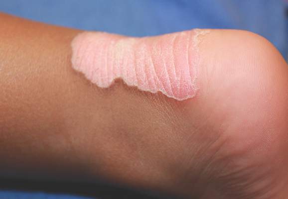
A 3-year-old girl presented with an erythematous, hypopigmented, scaling plaque on the posterior aspect of the left ankle surrounded by a hard rim. The plaque was first noted at 12 months of age and had slowly enlarged as the patient grew. Six months prior, a biopsy from the center of the lesion performed at another facility demonstrated a papulosquamous dermatitis.
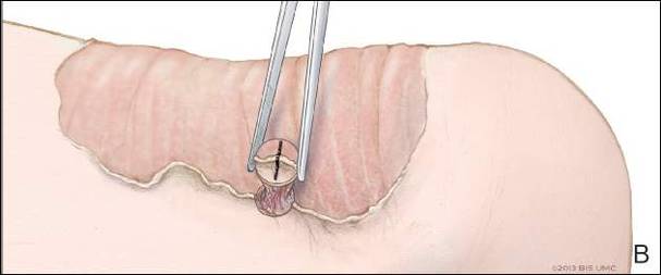
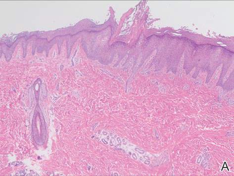
Physical examination revealed a lesion that was 4.2-cm long, 2.2-cm wide at the superior pole, and 3.5-cm wide at the inferior pole (Figure 1). A line was drawn with a skin marker perpendicular to the rim of the lesion (Figure 2A) and a 6-mm punch biopsy was performed, centered at the intersection of the drawn line and the cornoid lamella (Figure 2B). The tissue was then bisected at the bedside along the skin marker line with a #15 blade (Figure 2C) and submitted in formalin for histologic processing. Histologic examination revealed an invagination of the epidermis producing a tier of parakeratotic cells with its apex pointed away from the center of the lesion. Dyskeratotic cells were noted at the base of the parakeratosis (Figure 3). Verrucous hyperplasia was present in the central portion of the specimen adjacent to the cornoid lamella. Based on these histopathologic findings, the correct diagnosis of PM was made.
Figure 1. An erythematous scaling patch surrounded by a thin rim (cornoid lamella) typical of porokeratosis of Mibelli.
Figure 2. A skin marker was used to draw a line perpendicular to the cornoid lamella at the end of the lesion (A). After local anesthesia was administered, a 6-mm punch was centered at the intersection of the drawn line and the cornoid lamella (B). The punch specimen was bisected with a #15 blade along the line that was previously drawn (C). Illustrations by Kyle Cunningham, University of Mississippi Medical Center (Jackson, Mississippi).
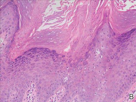
Figure 3. Histology revealed a broad cornoid lamella that erupted from a depression within the epidermis (A) (H&E, original magnification ×100). A close-up view of the cornoid lamella showed dyskeratotic cells beneath the column of parakeratosis (B)(H&E, original magnification ×400).