While the diagnostic challenges of photodermatoses are multiple, one thing is certain, a proper and rapid diagnosis is essential to optimizing patient management and outcomes, according to Dr. Vincent DeLeo.
The diagnostic challenges of photodermatoses include increasing clinical suspicion to recognize the less common forms, ruling out dermatologic mimics in the differential diagnosis, and identifying etiology to guide therapy and speed patient recovery, Dr. DeLeo noted at the annual Hawaii Dermatology Seminar sponsored by Skin Disease Education Foundation.
Photodermatosis incidence varies from the relatively common to the relatively rare, he said. Specific presentations include porphyria, idiopathic photodermatitis, polymorphic light eruption, solar urticaria, chronic actinic dermatitis, exogenous chemical photosensitivity, and photocontact dermatitis.
Consider age of onset, timing of the skin eruption, and occupation, medication, or plant exposures when taking patient history. Ask about products used for personal care, particularly any sunscreens or colognes. Clinical workup should include a review of systems that focuses on cutaneous and neurologic effects and a complete physical examination, said Dr. DeLeo, chairman of the department of dermatology at St. Luke's-Roosevelt Hospital Center and Beth Israel Medical Center in New York.
Porphyrias present in two specific patterns - immediate with skin burning within a day of exposure or delayed with blistering, hirsutism, or hyperpigmentation. With an immediate form, such as erythropoietic protoporphyria (EPP), rule out solar urticaria and lupus erythematosus, Dr. DeLeo suggested.
With a delayed presentation, such as porphyria cutanea tarda (PCT), consider pseudo-PCT and epidermolysis bullosa acquisita in your differential diagnosis, he said. A blood test for plasma porphyrins is usually diagnostic in all porphyrias, but a red blood cell porphyrin assay will diagnose EPP and a 24-hour urine assay will diagnose PCT.
Among the sporadic presentations of PCT, it is important to check for a diagnosis of hemochromatosis early to prevent serious tissue damage, especially dysfunction of the liver, Dr. DeLeo said. A test for the culprit gene mutations can be ordered from any major diagnostic laboratory.
Idiopathic photodermatoses include polymorphous light eruption (PMLE), chronic actinic dermatitis, and solar urticaria. Dermatologists are most likely to encounter PMLE, the most common form of photosensitivity, Dr. DeLeo said.
PMLE tends to arise in young adults and in women more than men. Symptoms usually occur within days of exposure - during springtime or while on vacation, for example. Look for a symmetrical distribution on exposed areas only. The histologic differential with suspected PMLE includes lupus erythematosus, lymphocytic infiltrate of Jessner, and cutaneous lymphoid hyperplasia.
Solar urticaria also occurs more often in women than in men. A sudden onset of erythema, edema, urticaria, and pruritus in exposed areas is characteristic. Systemic involvement is rare, Dr. DeLeo said. Careful photo provocation can aid in the definitive diagnosis of this condition. Treatment options include H1 antihistamines and proper photoprotection.
Chronic actinic dermatitis, on the other hand, is more persistent and not related to a specific acute exposure. Patients typically present with patchy eczema and may have erythroderma or lymphoma-like plaques in sun-exposed areas.
This form of photosensitivity is much more common in men and tends to present in older patients, at a mean age of 65 years, Dr. DeLeo said. Rigorous photoprotection is warranted. Consider patch testing and photopatch testing to rule out underlying contact or photocontact allergy. PUVA or immunosuppressants can be used in severe cases.
Some patients can experience photoirritant contact dermatitis from plants. A number of plants can cause this, including celery, dill, lemon, lime, and St. John's wort, he concluded.
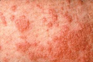
Dermatologists are most likely to encounter PMLE (shown above). It tends to arise in young adults and in women more than men. Symptoms usually occur within days of exposure. (Photo Courtesy: Dr. Vincent DeLeo)
Dr. DeLeo disclosed being a consultant for Neutrogena, Schering-Plough, L'Oreal, Pfizer, Estée Lauder, Orfagen, Goodyear, and Limited Brands. SDEF and this news organization are owned by Elsevier.