Case Reports

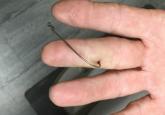
Don’t Get Hung Up on Fishhooks: A Guide to Fishhook Removal
In the sport of fishing, barbed fishhooks often are used for their effectiveness in maintaining the fish on the hook once it is caught.
From the Section of Dermatology, Department of Medicine, University of Chicago, Illinois.
The authors report no conflict of interest.
Correspondence: Clifford Hsieh, MD, 5841 S Maryland Ave, MC 5067, Chicago, IL 60637 (clifford.hsieh@uchospitals.edu).

Injuries from sea urchin spines are commonly seen in coastal regions with high levels of participation in water activities. Although these injuries may seem minor, the consequences vary based on the location of the injury. Sea urchin spine injuries may cause arthritis and synovitis from spines in the joints. Nonjoint injuries have been reported, and dermatologic aspects of sea urchin spine injuries rarely have been discussed. We present a case of a patient with sea urchin spines embedded in the thigh who subsequently developed painful skin nodules. Tissue from the site of the injury demonstrated foreign-body type granulomas. Following the removal of the spines and granulomatous tissue, the patient experienced resolution of the nodules and associated pain. Extraction of sea urchin spines can attenuate the pain and decrease the likelihood of granuloma formation, infection, and long-term sequelae.
Practice Points
Sea urchin injuries are commonly seen in coastal regions near both warm and cold salt water with frequent recreational water activities or fishing. Sea urchins belong to the class Echinoidea with approximately 600 species, of which roughly 80 are poisonous to humans.1,2 When a human comes in contact with a sea urchin, the spines of the sea urchin (made of calcium carbonate) can penetrate the skin and break off from the sea urchin, becoming embedded in the skin. Injuries from sea urchin spines are most commonly seen on the hands and feet, as the likelihood of contact with a sea urchin is greater on these sites. The severity of sea urchin spine injuries can vary widely, from minimal local trauma and pain to arthritis, synovitis, and occasionally systemic illness.1,3 It is important to recognize the wide variety of responses to sea urchin spine injuries and the impact of prompt treatment. Many published reports on injuries from sea urchin spines describe arthritis and synovitis from spines in the joints.1,2,4-6 Fewer reports discuss nonjoint injuries and the dermatologic aspects of sea urchin spine injuries.3,7,8 We pre-sent a case of a patient with a puncture injury from sea urchin spines that resulted in painful granulomas.
A 29-year-old otherwise healthy man was referred to our dermatology clinic by the university student health center due to continued pain in the right thigh. Five weeks prior to presentation to the student health center, the patient had fallen on a sea urchin while snorkeling in Hawaii. Sea urchin spines became lodged in the right thigh, some of which were removed in a local medical clinic in Hawaii. He was given oral antibiotics prior to his return home. A plain film radiograph of the affected area ordered by the student health center showed several punctate and linear densities in the lateral aspect of the right mid thigh (Figure 1). These findings were consistent with sea urchin spines within the superficial soft tissues of the lateral thigh.
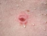
At the time of presentation to our dermatology clinic, the patient reported sharp intermittent pain localized to the right thigh. The patient denied any fever, chills, or pain in the joints. On physical examination, there were several firm nodules on the right thigh, ranging from 4 to 20 mm in diameter (Figure 2). The nodules were tender to palpation with some surrounding edema. Drainage was not noted. Several scars were visible at sites of the original puncture injuries and removal of the spines.
Two 6-mm punch biopsies were performed on representative nodules on the right thigh for histopathologic examination. Along with the biopsy tissue, firm, brown-black, linear foreign bodies consistent with sea urchin spines were extracted with forceps (Figure 3). Histopathologic examination revealed a dense, diffuse, mixed inflammatory cell infiltrate in the dermis predominantly composed of lymphocytes, histiocytes, and numerous eosinophils. Proliferation of small vessels was noted. In one of the biopsies, small fragments of necrotic tissue were present. These findings were consistent with granulomatous inflammation and granulation tissue due to a foreign body.
At the time of suture removal 2 weeks later, the biopsied areas were well healed with minimal erythema. The patient reported decreased pain in the involved areas. He was not seen in clinic again due to resolution of the nodules and associated pain.

In the sport of fishing, barbed fishhooks often are used for their effectiveness in maintaining the fish on the hook once it is caught.

Fish infrequently are aggressive toward humans. Rare acts of aggression often appear to be accidental, in self-defense, or for territorial...

Ethnic diversity in the United States has increased the presence of a variety of exotic foods. A lack of knowledge about certain foods, including...
