From the Journals

Nailfold analysis can predict cardiopulmonary complications in systemic sclerosis

View on the News

NVC’s role shifts from diagnostic to prognostic

Systemic sclerosis is a profoundly heterogeneous disorder, with the overall prevalence of major organ-specific manifestations, such as pulmonary arterial hypertension (PAH), broadly adhering to a 15% rule. As such, the majority of patients with SSc will not develop any given organ-specific complication. The major challenge for clinicians during the early stages of the disease is predicting the future occurrence of potentially life-threatening organ-specific manifestations, such as PAH.

The complementary association of nailfold videocapillaroscopy changes and autoantibody profile in predicting cardiopulmonary involvement reported by Dr. Markusse and her colleagues is novel, but otherwise supports the findings of previous cross-sectional studies identifying associations between advanced NVC changes and SSc complications, such as digital ischemic lesions and PAH. These studies provide intriguing insight into the relationship between the evolution of microangiopathy and the emergence of organ-specific manifestations of SSc, but also represent a shift in focus from the diagnostic to the prognostic utility of NVC in SSc.

There is potential clinical utility in these observations that has yet to be unlocked fully; particularly should the predictive value and timing of NVC progression be further characterized in longitudinal studies better defining the natural history of SSc organ-specific manifestations. If evolving NVC changes (in high-risk serological subgroups) are shown to pre-date the emergence of overt organ-specific manifestations of SSc, then we might be provided with a window of opportunity for escalation of therapy with treatments targeting endothelial function (such as phosphodiesterase inhibitors and/or endothelin receptor antagonists) and/or possible immunomodulatory approaches. This could potentially usher in a new era of preventive disease-modifying therapeutic intervention in SSc.

John D. Pauling, MD, PhD, is a consultant rheumatologist at the Royal National Hospital for Rheumatic Diseases, Bath, England, and Visiting Senior Lecturer in the department of pharmacy and pharmacology at the University of Bath. His commentary is derived from an editorial accompanying the study by Dr. Markusse and her associates (Rheumatology [Oxford]. 2016 Dec 30. doi: 10.1093/rheumatology/kew461). He disclosed having received grants and consultancy income from Actelion.


FROM RHEUMATOLOGY

Nailfold videocapillaroscopy can help to predict which patients with systemic sclerosis may develop serious cardiopulmonary complications, according to findings from a Dutch cross-sectional study.

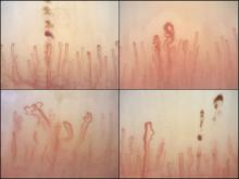
Nailfold capillaroscopy images show progressively worsening damage: microhemorrhage and hemosiderin (top left), an isolated dilated loop (top right), lower capillary density and microhemorrhage (lower right), and architectural derangement (lower left). ACR Copyright 2017

Nailfold capillaroscopy images show progressively worsening damage: microhemorrhage and hemosiderin (top left), an isolated dilated loop (top right), lower capillary density and microhemorrhage (lower right), and architectural derangement (lower left).

For their research, Iris M. Markusse, MD, PhD, and her colleagues at Leiden (the Netherlands) University Medical Center collected data on nailfold videocapillaroscopy (NVC) patterns and SSc-specific autoantibodies from a cross section of 287 patients in an established SSc cohort (Rheumatology [Oxford]. 2016 Dec 10. doi: 10.1093/rheumatology/kew402).

All patients in the study had NVC pattern data as well as anti-extractable nuclear antigen (anti-ENA) antibodies. The mean age of the patients was 54 years; 82% were female, and median disease duration was 3 years. Just over half the cohort had interstitial lung disease, and 16% had pulmonary artery hypertension.

Among the anti-ENA autoantibody subtypes, anti-ACA was seen in 37% of patients, anti-Scl-70 in 24%, anti-RNP in 9%, and anti-RNAPIII in 5%; other subtypes were rarer. SSc-specific NVC patterns were seen in 88% of patients, with 10% of the cohort showing an early (less severe microangiopathy) pattern, 42% an active pattern, and 36% a late pattern.

One of the study’s objectives was to determine whether one or more mechanisms was responsible for both autoantibody production and the microangiopathy seen in SSc.

If a joint mechanism is implicated, “more severe NVC patterns would be determined in patients with autoantibodies (such as anti-Scl-70 and anti-RNAPIII) that are associated with more severe disease,” wrote Dr. Markusse and her colleagues. “On the other hand, if specific autoantibodies and stage of microangiopathy reflect different processes in the disease, a combination of autoantibody status and NVC could be helpful for identifying patients at highest risk for cardiopulmonary involvement.”

The investigators reported finding a similar distribution of NVC abnormalities across the major SSc autoantibody subtypes (except for anti–RNP-positive patients), suggesting that combinations of the two variables would be most predictive of cardiopulmonary involvement. More severe NVC patterns were associated with a higher risk of cardiopulmonary involvement, independent of the presence of a specific autoantibody.

Notably, the researchers wrote, “prevalence of ILD [interstitial lung disease] is generally lower among ACA-positive patients. According to our data, even among ACA-positive patients there was a trend for more ILD being associated with more severe NVC patterns (OR = 1.33).”

A similar pattern was seen for pulmonary artery hypertension. “Based on anti-RNP and anti-RNAPIII positivity, patients did not have an increased risk of a [systolic pulmonary artery pressure] greater than 35 mm Hg; however, with a severe NVC pattern, this risk was significantly increased (OR = 2.33).”

The investigators cautioned that their findings should be confirmed in larger cohorts. The study by Dr. Markusse and her colleagues was conducted without outside funding, though manufacturers donated diagnostic antibody tests. One of the 11 study coauthors disclosed receiving financial support from Actelion.

Recommended Reading

Tips on how to differentiate CLE from dermatomyositis
MDedge Dermatology
Be alert for dermatomyositis without muscle disease
MDedge Dermatology
Anticipate, treat GI issues in scleroderma
MDedge Dermatology
Tryptase gene variant linked to GI, joint, and skin symptoms
MDedge Dermatology
ADDRESS II study: Atacicept shows promise for SLE
MDedge Dermatology
Myeloablative HSCT bests IV CYC for systemic scleroderma
MDedge Dermatology
Low-dose IL-2 shows promise for refractory lupus
MDedge Dermatology
Mycophenolate puts lupus patients in remission faster than azathioprine
MDedge Dermatology
Prenatal exposure to hydroxychloroquine cuts risk of neonatal cutaneous lupus
MDedge Dermatology
Lupus patients may be shortchanged on lipid management
MDedge Dermatology