Dr. Wagener is from Aesthetic Surgery Associates, Allentown, Pennsylvania. Dr. Laskas is from Dermatology Limited, Media, Pennsylvania. Drs. Purcell and Ermolovich are from Advanced Dermatology Associates, Allentown.
The authors report no conflict of interest.
Correspondence: Marie Wagener, DO, Aesthetic Surgery Associates, Integrated Health Campus, 250 Centronia Rd, Ste 301, Allentown, PA 18104 (m.lewars@gmail.com).
Muckle-Wells syndrome (MWS) is a rare disorder inherited in an autosomal-dominant fashion that belongs to a group of hereditary periodic fever syndromes. It specifically belongs to the cryopyrin-associated periodic syndromes (CAPSs) in which there is a mutation in the NLRP3 (NLR family pyrin domain containing 3) gene that leads to overproduction of IL-1β, the source of the multisystem inflammatory symptoms. Muckle-Wells syndrome is characterized by a recurrent urticarial eruption that is associated with episodic fever, myalgia, arthralgia, malaise, progressive sensorineural hearing loss, and amyloid nephropathy (the most severe complication). Basal cell nevus syndrome (BCNS), or Gorlin syndrome, is a rare, autosomal-dominant inherited genodermatosis linked to a mutation in the PTCH1 (patched 1) gene and is characterized by a broad range of anomalies. We report the case of a patient with MWS and BCNS in whom basal cell carcinoma (BCC) treatment was complicated by symptoms of MWS.
An urticarial rash occurring in childhood with symptoms of fever, joint pain, and swelling along with visual symptoms should prompt consideration of a cryopyrin-associated periodic syndrome.
Histopathology shows a dermal neutrophilic infiltrate that tends to be perivascular and also may be perieccrine. This atypical urticaria contrasts with the typical lymphocytic and eosinophilic infiltrate seen in classic urticaria.
References
Muckle-Wells syndrome (MWS) was first described in 1962 and is part of a broad category of hereditary periodic fever syndromes that include the autoinflammatory syndromes and the cryopyrin-associated periodic syndromes (CAPSs). Unlike autoimmune diseases, autoinflammatory syndromes are not associated with antigen-specific T-cell responses or high titers of autoantibodies but are related to disorders of the innate immune system. Basal cell nevus syndrome (BCNS), or Gorlin syndrome, is a rare genodermatosis inherited in an autosomal-dominant fashion that is characterized by a broad range of anomalies. Most notable is the early and strong predisposition to develop several to hundreds of basal cell carcinomas (BCCs). Classic clinical features of MWS and a thorough history and physical examination can assist in the diagnosis of this rare entity.
Case Report
A 35-year-old woman with a history of BCNS, which had been diagnosed at 24 years of age based on the presence of more than 2 BCCs and a family history of BCNS in her mother, presented with intermittent pruritic urticaria on the chest and back, episodic fevers, associated joint pain and swelling that worsened several hours after exercise, headache, conjunctivitis, blurred vision, and severe debilitating fatigue that had been present since childhood. The symptoms had progressively worsened with age and symptom-free intervals became shorter. She was diagnosed by her rheumatologist with biopsy-proven MWS and a positive NLRP3 (NLR family pyrin domain containing 3) gene mutation at 29 years of age. She was treated unsuccessfully with prednisone and antihistamines and entered a trial with anakinra. She showed improvement for 2 weeks but developed severe swelling and erythema at the injection sites at week 3, along with large leathery patches on the legs and difficulty ambulating.
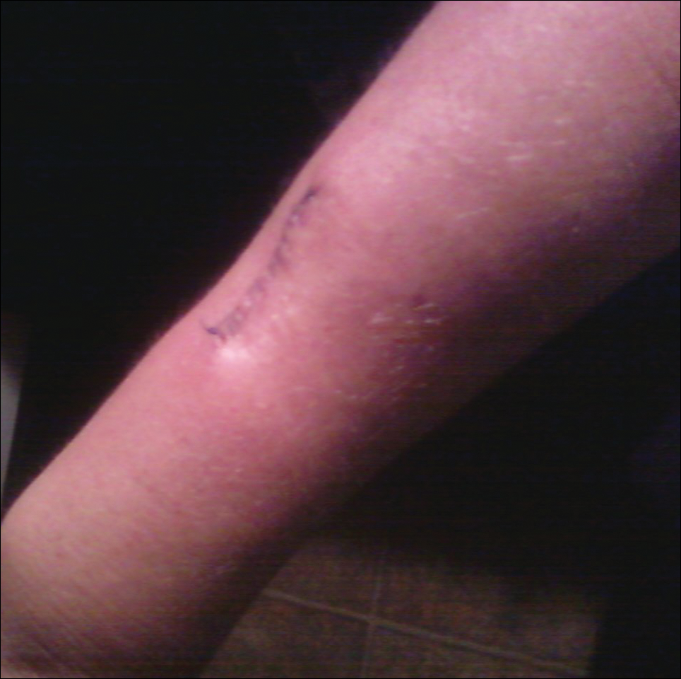
The patient subsequently underwent excision of her BCCs and reported each site became erythematous, edematous, warm, and painful 6 hours after excision, which lasted for hours to days (Figures 1–3). After the first excision on the right forearm, she was seen in the emergency department, started on intravenous antibiotics and prednisone, and kept overnight in the hospital. She was discharged the following day and the edema in the right forearm subsided over several days. Bacterial culture and laboratory evaluation for infection were negative after the first excision on the right forearm. Because of the symptoms she experienced following this excision, she was referred to the plastic surgery department for excision followed by postoperative monitoring in the hospital. The patient continued to undergo excisions for BCCs and developed more severe symptoms including erythema, edema, warmth, and tenderness at the surrounding sites. Once again, the excision sites were cultured and laboratory work to rule out infection was ordered with a negative result. After several excisions and subsequent clinical findings, the patients’ symptoms were deemed consistent with MWS and not a result of infectious etiology. A diagnosis of MWS and BCNS with exacerbation of MWS with surgical procedures was made.
Figure 1. Erythema, edema, warmth, and tenderness surrounding the excision site on the right forearm 6 hours after basal cell carcinoma excision.
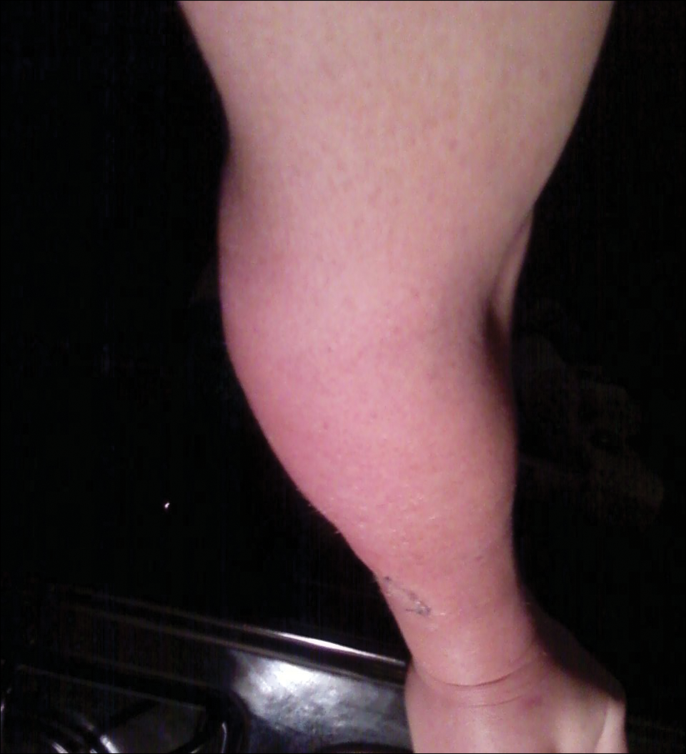
Figure 2. Erythema, edema, warmth, and tenderness surrounding the excision site on the right arm spreading distally to include the right wrist 24 hours after basal cell carcinoma excision.
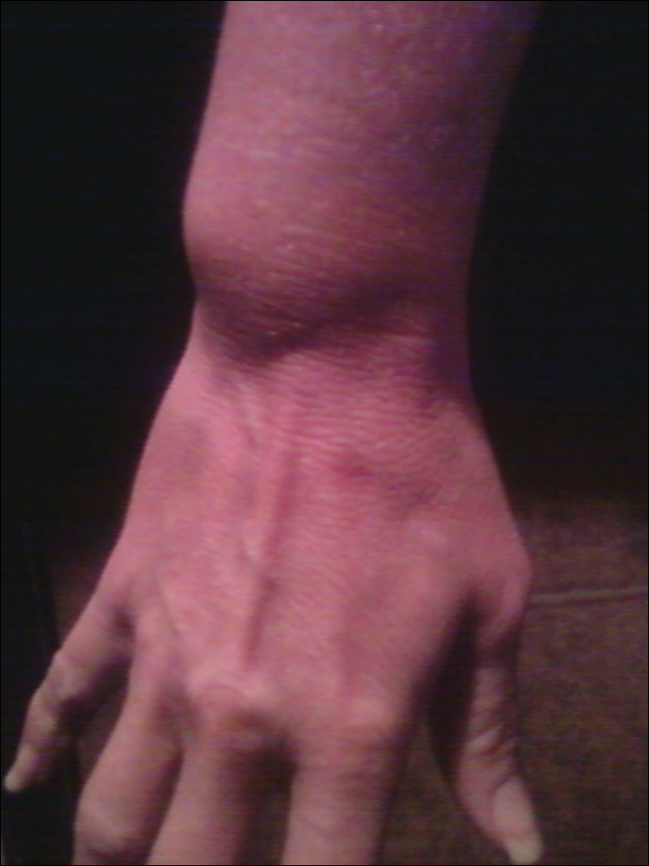
Figure 3. Erythema, edema, warmth, and tenderness on the right wrist distal from the excision site 3 days after basal cell carcinoma excision.
The patient has continued therapy with rilonacept for MWS, which is managed by her rheumatologist. She has tolerated rilonacept without adverse effects and has experienced a reduction in symptoms that has enhanced her quality of life and allows for further treatment of her BCNS. Her dermatologist (J.W.L.) has been treating her BCCs with vismodegib, but treatment has been sporadic due to muscle cramping after 7 days of therapy. She reported subjective improvement to her dermatologist and has tried alternating 7 days on and 7 days off vismodegib. The muscle cramping still has limited her treatment with this regimen, and she is currently on a trial of 3 days on, 4 days off per week.