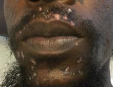
Cutaneous lupus erythematosus can be classified into acute, subacute, and chronic lesions. Chronic cutaneous lupus, or discoid lupus erythematosus (DLE), may occur independently of or in combination with systemic lupus erythematosus (SLE). They are one of the more common skin presentations seen in lupus. Young adults are typically affected, with a female-to-male ratio of 2:1. Progression from DLE to SLE is uncommon. However, patients with SLE will frequently develop discoid lesions.
Lesions generally occur on the head and neck, with scalp and ears (conchal bowls) frequently affected. DLE lesions often begin as erythematous papules or plaques that may become scaly and heal with atrophy, scarring and dyspigmentation (often central hypopigmentation with peripheral hyperpigmentation). Follicular plugging is often seen in lesions. Erosions may occur. A small percentage of patients may have mucosal involvement, including the lips. Sun exposure may have a role in the development of lesions, although lesions may also occur in non–sun exposed areas. Less commonly, DLE may be generalized and involve the trunk and extremities, in addition to the head and neck. Scarring alopecia can be present on the scalp. Scarring may become disfiguring.The differential diagnosis includes: subacute cutaneous lupus, lichen planus, seborrheic dermatitis, Jessner’s lymphocytic infiltrate, polymorphous light eruption, rosacea, granuloma faciale, and sarcoidosis. Histology of DLE may reveal hyperkeratosis, a thin epidermis with effacement of the rete ridges, a lichenoid and vacuolar interface dermatitis, and follicular plugging. Damaged keratinocytes called colloid bodies may be present. Increased mucin and thickening of the basement membrane are commonly seen. Active lesions will exhibit more of an inflammatory infiltrate. Direct immunofluorescence of lesional skin is positive in more than 75% of cases.
Treatment includes sunscreen and avoidance of sun exposure. Potent or superpotent topical corticosteroids, as well as lesional injections of triamcinolone are helpful. Although, generally, it is not advised to use a high-potency steroid on the face, it can be helpful in DLE. Application should be limited to affected areas for short periods of time, with frequent monitoring for possible side effects. Topical calcineurin inhibitors can be used in addition to topical corticosteroids. If systemic treatment is indicated, hydroxychloroquine is first line. Short-term oral corticosteroid treatment can be used while transitioning to other systemic medications. Our patient had negative serologies and responded to high-dose topical steroids with complete clearing of cutaneous lesions.This case and the photo were submitted by Dr. Bilu Martin.
Dr. Bilu Martin is a board-certified dermatologist in private practice at Premier Dermatology, MD, in Aventura, Fla. More diagnostic cases are available at edermatologynews.com. To submit a case for possible publication, send an email to dermnews@frontlinemedcom.com.


