Case Reports

Eruptive Vellus Hair Cysts in Identical Triplets With Dermoscopic Findings

Author and Disclosure Information

Eruptive vellus hair cysts (EVHCs) are asymptomatic, follicular, flesh-colored to hyperpigmented papules that are typically located on the chest and extremities with an unclear inheritance pattern and pathogenesis. We report a case of EVHCs in 4-year-old identical triplet girls. Our aim is to present another rare case of autosomal-dominant inheritance of EVHCs and to emphasize the utility of dermoscopy as a diagnostic aid for EVHCs that may be misdiagnosed clinically.

Practice Points

  • Eruptive vellus hair cysts (EVHCs) are 1- to 3-mm round, dome-shaped, flesh-colored, asymptomatic, benign papules typically occurring on the chest and extremities.
  • Pathogenesis and inheritance are unclear. Although the majority of EVHC cases are sporadic, the strong influence of genes is indicated by numerous reports of families in whom 2 or more members were affected.
  • Dermoscopy is a noninvasive diagnostic procedure that should be utilized to diagnose EVHCs in the pediatric population; specifically, EVHCs exhibit light yellow, homogenous, circular structures with a maroon or erythematous halo.
  • The main indication for treatment of EVHCs is cosmetic concern; however, one-quarter of cases may resolve spontaneously.


References

Case Report

Four-year-old identical triplet girls with numerous asymptomatic scattered papules on the chest of 4 months’ duration were referred to a dermatologist by their pediatrician for molluscum contagiosum. The patients’ father reported that there was no history of trauma, irritation, or manipulation to the affected area. Their medical history was notable for prematurity at 32 weeks’ gestation and congenital dermal melanocytosis. Family history was notable for their father having acne and similar papules on the chest during adolescence that resolved with isotretinoin therapy.

On physical examination there were multiple smooth, hyperpigmented to erythematous, comedonal, 1- to 2-mm papules dispersed on the anterior central chest of all 3 patients (Figure 1). Clinically, these lesions were fairly indistinguishable from other common dermatologic conditions such as acne or milia. Dermoscopic examination revealed homogenous yellow-white areas surrounded by light brown to erythematous halos (Figure 2). Histopathologic examination was not performed given the benign clinical diagnosis and avoidance of biopsy in pediatric populations. Based on dermoscopic features and history, a diagnosis of eruptive vellus hair cysts (EVHCs) in identical triplets was made.

Figure1

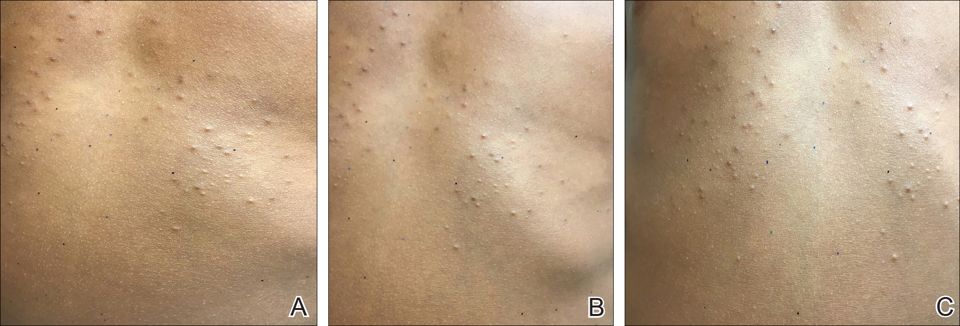
Figure 1. Scattered erythematous, flesh-colored papules on the central chest with flecks of glitter on each of the identical triplet girls (A–C).

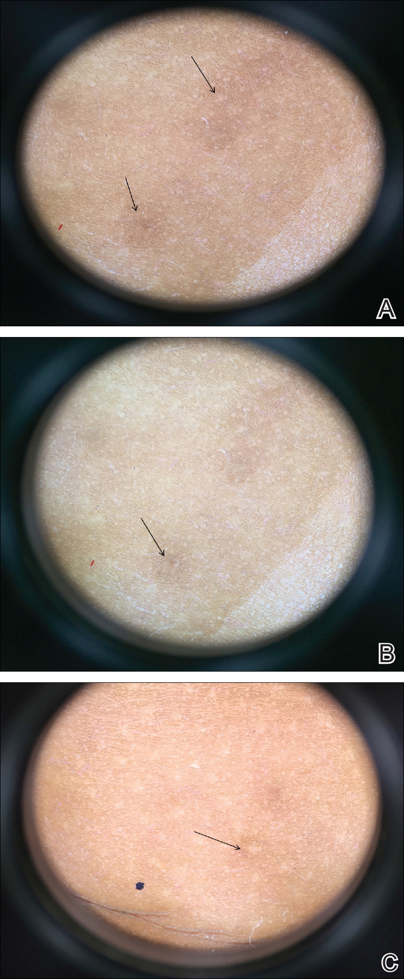
Figure 2. Dermoscopic findings of homogenous yellow-white areas surrounded by light brown to erythematous halos (black arrows)(A–C).

Pages

Recommended Reading

Longitudinal melanonychia: the good, the bad, and the confusing
MDedge Dermatology
Alopecia areata linked to mental health disorders
MDedge Dermatology
So it’s pediatric onychomycosis. Now what?
MDedge Dermatology
JAK inhibitors emerge as promising alopecia treatment
MDedge Dermatology
Expert shares laser hair removal clinical pearls
MDedge Dermatology
A new era arrives in alopecia areata therapy
MDedge Dermatology
Platelet-rich plasma injections yield substantial improvement in androgenetic alopecia
MDedge Dermatology
Diffuse Nonscarring Alopecia
MDedge Dermatology
Pigmented Pruritic Macules in the Genital Area
MDedge Dermatology
Frontal Fibrosing Alopecia: Cutaneous Associations in Women With Skin of Color
MDedge Dermatology