SAN DIEGO - A remedy that has been used for nearly 2 centuries is still a valuable adjuvant therapy for melanoma, according to Dr. Norman Brooks.
Zinc chloride paste not only penetrates and kills even thick melanomas, it appears to stimulate a strong immune reaction that decreases the likelihood of metastasis and recurrence, Dr. Brooks said at the meeting sponsored by the American Society for Mohs Surgery.
Dr. Norman Brooks
"I believe chemosurgery with zinc chloride paste, followed by wide excision, is a better way to remove a melanoma than fresh tissue surgery," said Dr. Brooks, a dermatologist in Encino, Calif., who specializes in skin cancers. "It not only kills the tumor locally but stimulates total body resistance and immunity, supporting the idea that surgery actually enhances melanoma tumor growth and metastatic spread. That’s why I believe it's so important to use zinc chloride when treating melanoma."
History of Zinc Chloride
Zinc chloride was first used as a cancer treatment in 1815. In 1858, a U.S. physician added a powdered form of the bloodroot plant (Sanguinaria canadensis), which contains the anticancer alkaloid sanguinarine; the compound was used to treat breast cancer. The current paste, formulated by Dr. Frederic E. Mohs, is a compound of naturally occurring ingredients: the mineral stibnite, which forms the inert paste-like vehicle; powered root of the bloodroot plant, which has been shown to induce apoptosis in several types of cancer; and a saturated zinc chloride solution, which destructively penetrates tissue.
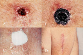 Photo courtesy Dr. Norman Brooks
(A) Saucerized fresh-tissue biopsy site of a level IV 1.8-mm thick amelanotic melanoma on the left arm of a 67-year-old male. (B) Preoperative treatment of site with zinc chloride fixative paste. (C) Occlusive dressing to hold paste in place for 24 hours. (D) Subsequent conventional fresh-tissue excision of a deep wide margin.
A 1997 study explored the idea that surgery can liberate atypical melanocytes and predispose a patient to metastatic disease, Dr. Brooks said (Dermatol. Surg. 1997;23:1043-6). The epidemiologic study of 1,224 primary melanomas concluded that surgery of primary melanoma may enhance tumor growth at metastatic sites.
Chemosurgery with zinc chloride paste, therefore, helps avoid this phenomenon, Dr. Brooks said. The paste not only destroys tissue where it is applied, it also fixes the tissue into a firm mass. A delayed excision allows the specimen to be removed en bloc and examined histopathologically to determine margins. However, leaving a portion of the fixed tissue in place for several more days causes a strong vaccine-like reaction on the skin surface, indicating, according to Dr. Brooks, the beginning of a systemic immunologic response.
The Evidence
According to Dr. Brooks, he and his colleagues published the only piece of objective evidence on this phenomenon in 1998 (Dermatol. Surg. 1998;24:1021-5).
The study involved mice that were injected with two different melanoma types: a poorly immunogenic (B16) type and a more immunogenic type (K1735p). The resultant tumors were freshly excised in some mice, or excised 24 hours after the application of zinc chloride paste. A week later, all the mice received a second injection of melanoma cells at a different site. K1735p tumors developed at the challenge sites in significantly fewer of the mice pretreated with zinc chloride (32% vs. 69%, respectively). The paste did not alter the development of B16 melanomas. The authors concluded: "Zinc chloride fixation of the more immunogenic K1735p melanoma increased resistance to subsequent tumor challenge, suggesting that zinc chloride fixative paste acts as an immune adjuvant."
Treatment Application
At the meeting, Dr. Brooks presented his most recent article describing a simplified method for using the paste (Dermatol. Surg. 2010;36:237-40).
His process begins with a fresh tissue biopsy to confirm melanoma. He recommended a shaved saucerized excision with a margin of 1-2 mm beyond the suspected tumor edge carried at least through the mid-dermis, or below the suspected thickness of the lesion.
After melanoma is histopathologically confirmed, he applies the Mohs paste. Since the compound will not penetrate an intact keratin layer, it must only be applied to the excised area. "If the biopsied area has developed a crust or eschar, this must also be removed," Dr. Brooks said.
He applies a 1-mm thick layer of paste with a cotton-tipped swab. More can be applied for thicker lesions, but less if the lesion overlies an important anatomic structure. If the biopsy is a small excisional one, a swab of 50% bi- or trichloracetic acid can remove the outer keratin layer and allow the paste to penetrate.