Make the Diagnosis

A woman with scaling, and painful, crusted, erythematous papules and pustules on her face

A 78-year-old White female presented with a 1 year history of scaling, and painful, crusted, erythematous papules and pustules on the right lateral cheek, preauricular region, and ear. She had multiple treatments in the past, including cephalexin, prednisone, topical steroids, topical antibiotics, and topical ivermectin. The patient reported that topical medications seemed to aggravate the eruption. Biopsies were performed and a KOH prep was performed.

What's your diagnosis?

Granulomatous rosacea

Erosive pustular dermatosis

Demodex folliculitis

Pustular psoriasis

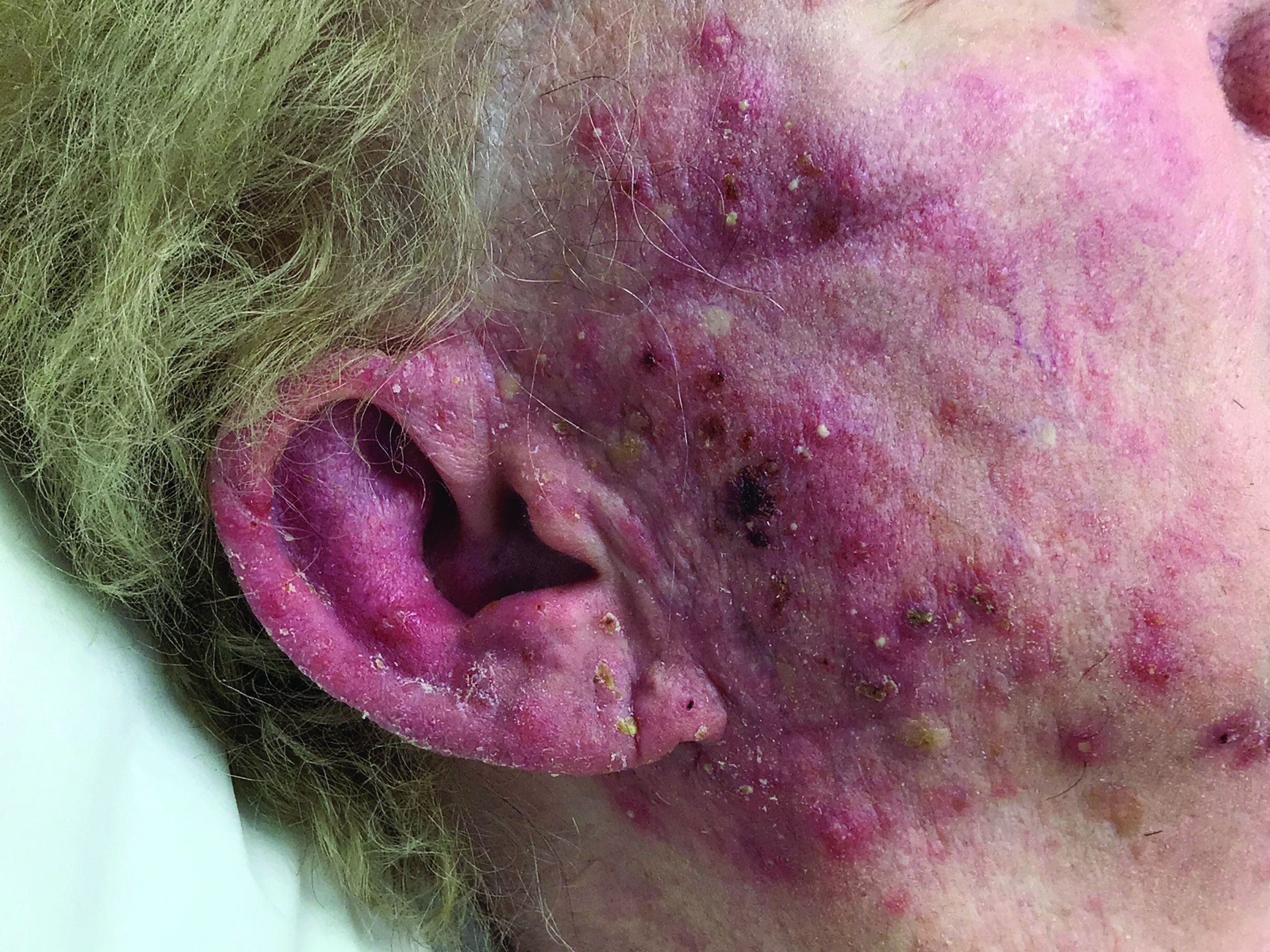
Biopsy for this patient revealed folliculitis with Demodex mites visualized on histology. Direct immunofluorescence was negative. A KOH preparation was performed and was positive for large numbers of Demodex. Bacterial cultures were negative. The patient was started on a course of submicrobial doxycycline and ivermectin and showed marked improvement 1 month following treatment.

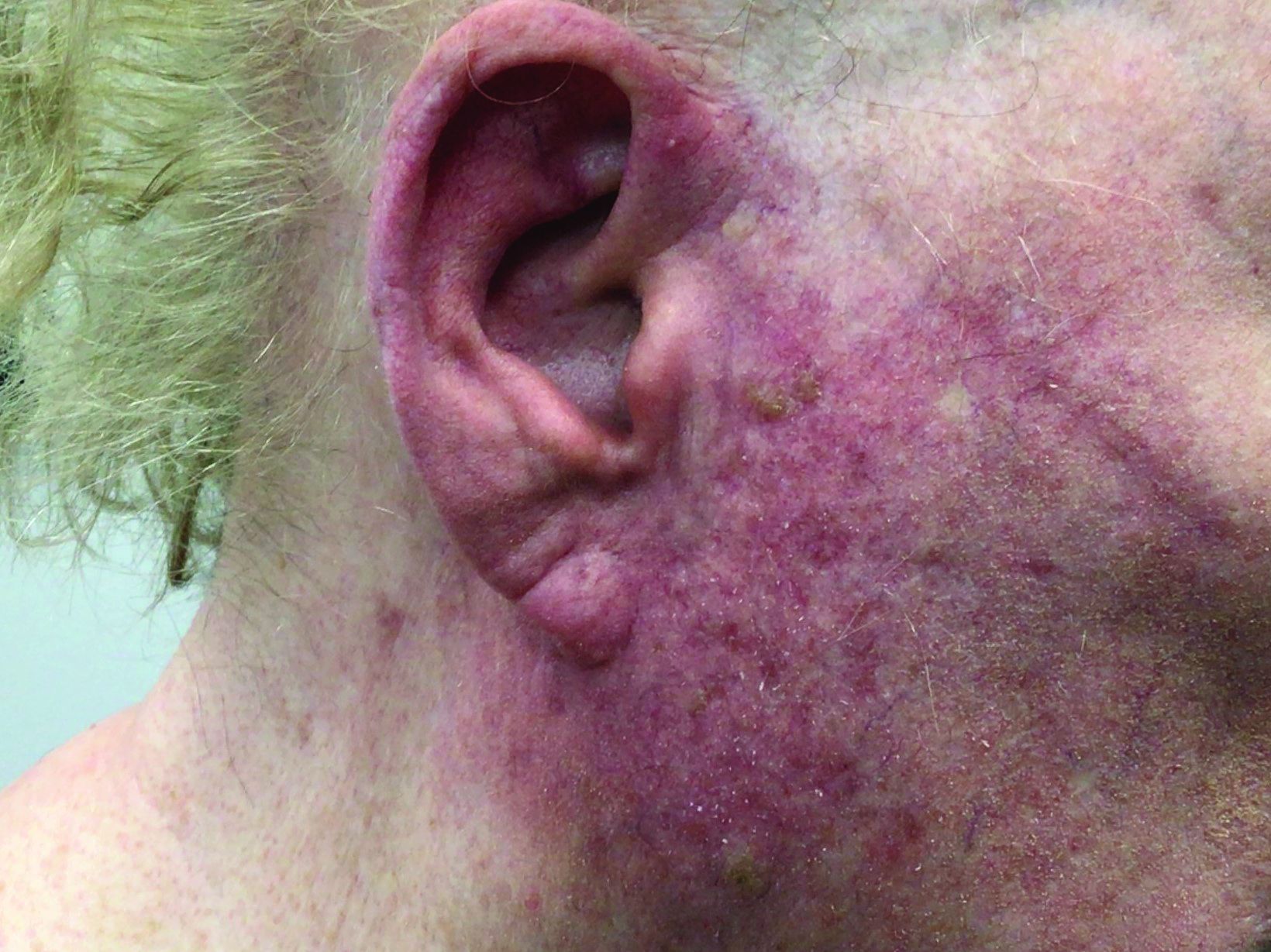
Demodex folliculorum and Demodex brevis (collectively referred to as Demodex) are microscopic parasitic mites that commonly live on human skin.1 Typically, the mite remains asymptomatic. However, in higher numbers, the infestation may cause dermatoses, called demodicosis. Demodex folliculitis is caused by high numbers of Demodex folliculorum, which lives in follicles and sebaceous glands. Lesions often present as itchy papules, pustules, and erythematous scaling on the face, ears, and scalp. Blepharitis may be present. Demodex folliculitis is more common in immunocompromised patients.2

Demodex may have a causative role in rosacea and present similarly, with a key difference being that Demodex-type rosacea is more scaly/dry and pustular than common rosacea.1 In Demodex folliculitis, bacterial cultures are often negative. A skin scraping for KOH will reveal increased mite colonization. The Demodex mite may also be seen in histologic slides.

Treatment of Demodex folliculitis includes crotamiton cream, permethrin cream, oral tetracyclines, topical or systemic metronidazole, and topical or oral ivermectin.

This case and photos were submitted by Susannah McClain, MD, Three Rivers Dermatology, Pittsburgh.

References

1. Rather PA and Hassan I. Indian J Dermatol. 2014 Jan;59(1):60-6.

2. Bachmeyer C and Moreno-Sabater A. CMAJ. 2017 Jun 26;189(25):E865.

Recommended Reading

Cutaneous Manifestations of COVID-19: Characteristics, Pathogenesis, and the Role of Dermatology in the Pandemic
MDedge Dermatology
Phage-targeting PCR test picks up early Lyme disease
MDedge Dermatology
Ten reasons airborne transmission of SARS-CoV-2 appears airtight
MDedge Dermatology
Vaccinating homebound patients is an uphill battle
MDedge Dermatology
Tinea Incognito Mimicking Pustular Psoriasis in a Patient With Psoriasis and Cushing Syndrome
MDedge Dermatology
Head to Toe: Recommendations for Physician Head and Shoe Coverings to Limit COVID-19 Transmission
MDedge Dermatology
Genital Primary Herpetic Infection With Concurrent Hepatitis in an Infant
MDedge Dermatology
Mohs Micrographic Surgery During the COVID-19 Pandemic: Considering the Patient Perspective
MDedge Dermatology
What’s Eating You? Culex Mosquitoes and West Nile Virus
MDedge Dermatology
CDC recommends use of Pfizer’s COVID vaccine in 12- to 15-year-olds
MDedge Dermatology