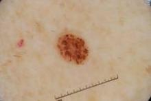
Theories of how nevi develop have received little attention in the last 100 years, but that appears to be changing, according to Dr. Ashfaq A. Marghoob.
Dr. Marghoob, a dermatologist at Memorial Sloan-Kettering Cancer Center in New York, discussed what is currently known about nevogenesis. "Since nevi are associated with an increased risk of melanoma, understanding nevogenesis may help to unravel some of the mysteries of melanomagenesis," he noted at the Hawaii Dermatology Seminar sponsored by Skin Disease Education Foundation (SDEF).
German physician Paul Gerson Unna proposed the "Abtropfung" theory of nevogenesis in 1893. The theory holds that melanocytic nevus cells develop in the epidermis and drop off to the dermis over time. "For almost a century this theory of nevogenesis was accepted as truth and remained uncontested," Dr. Marghoob said.
During the past few decades, however, newly acquired observations from histopathology and embryogenesis have led some researchers to question the validity of the "Abtropfung" theory in favor of the "Hochsteigerung" theory.
Dr. Stewart F. Cramer, a pathologist, proposed the latter in 1984. It essentially is the reverse of the Abtropfung theory, meaning that nevus cells migrate from the dermis to the epidermis.
For example, in one histopathology study, no child younger than 10 had a purely junctional nevus, 52% had compound nevi, and 48% had dermal nevi (Am. J. Dermatopathol. 1998;20:135-9). In patients older than age 60, 12% had junctional nevi, 23% had compound nevi, and 65% had dermal nevi. Had the Abtropfung theory been correct, most childhood nevi would be junctional, and most nevi in late adult life would be intradermal. The researchers said their findings better fit the theory of upward migration of nevus cells.
In another study, researchers reviewed all biopsy reports for junctional nevi from 2001 at the Penn State Milton S. Hershey Medical Center and found that these lesions occur with similar frequency in the young and in the elderly (J. Am. Acad. Dermatol 2007;56:825-7).
"However, new insights gained from the epidemiology of nevi, cross-sectional and longitudinal study of nevi, dermoscopy and confocal microscopy investigation of nevi, as well as the cellular and molecular study of nevi, brings into question the aforementioned theories," Dr. Marghoob says.
In fact, he added, there is insufficient evidence that either theory is the norm in postnatal life. More longitudinal studies are needed to determine nevus evolution.
Dr. Marghoob reported having no relevant conflicts of interest. SDEF and this news organization are owned by Elsevier.


