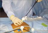
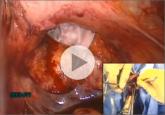
Dr. Kho: I infrequently perform supracervical hysterectomy, so almost all the hysterectomies I do are total hysterectomies. I remove the uterus through the vagina. In addition, because the size of the specimen frequently is too large to remove through a colpotomy intact, I morcellate the uterus manually with a scalpel using coring, wedge resection, and myomectomy. I find this to be an efficient and controlled method for tissue removal, with minimal tissue scattering. I also have begun to perform the same type of vaginal morcellation with the specimen enclosed in a bag.
That being said, the spread of occult malignancy has been reported after all types of morcellation—not just with power morcellation but also with vaginal and abdominal morcellation. So we are increasingly performing tissue extraction in an enclosed fashion using manual morcellation in a containment bag through a mini-laparotomy or posterior colpotomy to minimize the risk of leaving tissue fragments behind.
Dr. Wright: Although different methods of tissue extraction, including morcellation within a bag, are commonly discussed, data documenting the safety of these methods are extremely limited and patients should be counseled accordingly.
Similarly, the risk of adverse pathology increases substantially with age, and morcellation should be considered with great caution—if at all—in older women.
Given the risks associated with power morcellation, I try to avoid uterine disruption at the time of hysterectomy and perform either vaginal or minimally invasive total hysterectomy. In older women, because of the higher risk of underlying pathology, I prefer laparotomy if anatomic considerations preclude a vaginal or minimally invasive total hysterectomy. Younger women can be counseled about the risks and benefits of various routes of extraction. Patients with any suspicious findings during preoperative evaluation or surgery itself should have their uterus removed without disruption or fragmentation.
In regard to myomectomy specifically, a significant portion of the data we have on the risks of power morcellation derives from studies of hysterectomy. There are minimal data describing the risk of occult pathology at the time of minimally invasive myomectomy. Although younger patients likely are at relatively low risk for occult malignancy, they should be counseled that population-based estimates of cancer at the time of myomectomy are lacking.
Dr. Bradley: Since the controversy over morcellation arose, the Cleveland Clinic not only has banned the procedure but also removed all morcellators from its shelves, and it is unclear whether the option will be revisited after the FDA renders its final verdict. So my approach to tissue extraction is either vaginal morcellation or using a mini-laparotomy to remove the whole specimen intact or put it in a bag and morcellate it with a knife.
Case 1: A premenopausal patient scheduled for myomectomy
OBG Management: Let’s move on to a specific case. Let’s say the patient is a 35-year-old woman with a large fibroid, to be removed by myomectomy. How would you quantify her risk of occult malignancy? And what would preoperative assessment entail?
Dr. Iglesia: This patient’s risk of occult malignancy is low. I would obtain pelvic ultrasonography and endometrial biopsy, with cervical cytology included. Preoperative magnetic resonance imaging (MRI) would be indicated if there is a possibility that power morcellation will be performed. If power morcellation were selected, I would perform it using a bag.
Dr. Bradley: At the Cleveland Clinic, we now utilize the FDA risk estimates for occult malignancy of 1 in 300 to 1 in 350 women,1 and I counsel patients using these figures. In the past several years, we have begun to use MRI with and without contrast to determine the size, number, and location of the fibroids, to determine our surgical approach, and to guide our discussion with the patient of what we will be able to do—for example, laparoscopy versus laparotomy.
OBG Management: Would the FDA figures you give be applicable to a young woman such as this 35-year-old?
Dr. Bradley: We’re using those figures with all of our premenopausal patients.
OBG Management: And does the MRI pick up sarcomas?
Dr. Bradley: No imaging is 100% sensitive in detecting sarcoma. We do MRI, and if the fibroid has any areas of necrosis, irregularity, or poor tissue planes that would arouse our suspicion of adenomyoma or sarcoma, we perform the myomectomy via laparotomy. But as I mentioned earlier, we don’t use power morcellation at all anymore—so this patient you describe would likely undergo laparoscopic removal using a bag and a knife to extract it to the skin level.
Although every patient is different, in general, if we have a patient with a single large fibroid 10 cm or less in size, we try to remove it laparoscopically or with robot assistance rather than via laparotomy. We also perform endometrial biopsy.


