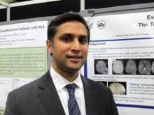
SAN DIEGO – Tube-shaped linear lesions seen on advanced MRI in people admitted to the emergency department for head injuries may not be diffuse axonal injury but rather evidence of bleeding due to mild traumatic brain injury, judging from preliminary results of a novel study.
"Not everything that we’re calling diffuse axonal injury is diffuse axonal injury," Dr. Gunjan Y. Parikh said in an interview during a poster session at the annual meeting of the American Academy of Neurology. "There may be, in fact, evidence under our noses of vascular injury. This has implications, and those patients should be followed very closely. If we can pinpoint that they’re a vascular injury in the acute setting, there are a lot of therapies [we can use] that target the vasculature."
Between October 2010 and October 2012 Dr. Parikh, a neuroimaging fellow at the National Institute of Neurological Disorders and Stroke, and his associates prospectively evaluated 256 patients enrolled in the Traumatic Head Injury Neuroimaging Classification (THINC) study who were admitted to the emergency department at Suburban Hospital in Bethesda, Md., and Washington (D.C.) Hospital Center after mild head injuries.
Administered by the Center for Neuroscience and Regenerative Medicine at Uniformed Services University in Bethesda, the THINC study protocol includes taking an MRI in subjects within 48 hours of presenting with acute head injury, with or without a positive diagnosis of concussion, and at up to three follow-up visits at 4, 30, and 90 days. The protocol includes T2-weighted MRI, fluid-attenuated inversion recovery (FLAIR), diffusion-weighted imaging (DWI), and three-dimensional tracking imaging (3-DTI).
The average age of the 256 patients was 50 years. Of these, 104 (41%) had imaging evidence of hemorrhage in the brain (67% reported loss of consciousness and 65% reported amnesia). These 104 patients underwent more detailed brain scans with advanced MRI within an average of 17 hours after the injury. This advanced imaging demonstrated that 20% of the 104 patients had microbleed lesions and 33% had tube-shaped linear lesions suggestive of vascular injury. Microbleeds were distributed throughout the brain, whereas linear lesions, which were found primarily in the anterior corona radiata, were more likely to be associated with injury to adjacent brain tissue.
"I was surprised that one-third of patients are having this linear type of lesion that we may think is vascular injury, and the majority of them – 91% – met the Glasgow Coma Scale criteria for mild TBI," Dr. Parikh commented. "These are patients who are usually sent home [after initial emergency department presentation]. I was also surprised to see so much evidence on other MRI sequences, proving that there is evidence of associated infarct or ischemia."
This type of analysis has not been done previously because "the logistical hurdles to imaging patients after any type of brain injury are so dramatic that it’s difficult to do this type of study," he said, emphasizing the preliminary nature of the work. "It was able to be done because of a unique collaboration between the National Institutes of Health (NIH) and the Department of Defense. If it weren’t for this collaboration, this would not have happened."
He noted that histopathological studies are "an important next step" to confirm the connection between the imaging markers and the pathology.
The study was supported by the NIH, the National Institute of Neurological Disorders and Stroke, and the Center for Neuroscience and Regenerative Medicine, a collaborative effort among the NIH, the Department of Defense, and Walter Reed National Military Medical Center, Bethesda. Dr. Parikh reported having no financial disclosures.


