MIAMI – Some hallmark signs of dermatologic problems in children – especially erythema and hyperpigmentation – often are less obvious in children with skin of color and can require more clinical detective work to diagnose.
Dr. Patricia A. Treadwell narrowed down the most likely dermatoses a pediatrician will encounter in this patient population at a pediatric update sponsored by Miami Children’s Hospital. Atopic and contact dermatitis, phytophotodermatitis, transient neonatal pustular melanosis, and neonatal lupus erythematosus are among the noteworthy clinical challenges, she said at the meeting.
"Children with increased pigmentation in their skin may end up testing your knowledge in terms of looking at their dermatitis and being able to diagnose that. Keep in mind it may be a little bit different in terms of the clinical presentation, but it’s important to identify it and start the proper treatment," said Dr. Treadwell, a pediatric dermatologist at Indiana University Health and Riley Hospital for Children in Indianapolis.
Overcoming this "masking" of a condition by skin pigmentation can be important, Dr. Treadwell said. She cited a patient born with a port-wine stain that went undiagnosed. The infant had subtle erythema and some asymmetry related to the overgrowth of the lesion. "This was not diagnosed based on the fact that the erythema was not apparent. The patient later developed a pyogenic granuloma, which is a complication that can be seen in patients with port-wine stains as they get older."

Dr. Patricia A. Treadwell
Erythema can be missed in children of color with atopic dermatitis as well. For this reason, atopic dermatitis may be underdiagnosed in this population overall, she said. Another challenge is the common practice of grading the severity of atopic dermatitis in lighter-skinned patients based on the degree of erythema. "In children with a fair amount of pigmentation in their skin, the erythema may not be noted and the severity will not be recognized."
Similarly, you might need a higher index of clinical suspicion to diagnose a child of color with contact dermatitis. Again, the erythema can be subtle. In contrast, "contact dermatitis can be very clear in a Caucasian patient. But the lesions are the same – linear, asymmetrical, and occurring on exposed areas," Dr. Treadwell said. Pruritus is common, and edema and swelling also occur. Watch for development of vesiculobullous lesions.
 Photos courtesy Dr. Patricia A. Treadwell
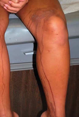
History taking was important to identify this phytophotodermatitis pattern of hyperpigmenation. Juice from a mango dripped on her leg before she went into the sun on vacation.
Phytophotodermatitis is another dermatologic condition that may require some additional detective work in children of color. Dr. Treadwell described a girl with a unique hyperpigmentation pattern on her legs and arms. She was referred following a vacation in Cancun, Mexico, with her family, and there was a concern about an autoimmune process. "They asked if she needed blood work. I said no, she was eating a mango and went out in the sun." Some of the mango juice splashed on her legs and arms.
Phytophotodermatitis occurs when furocoumarins from tropical fruit, citrus, celery, fennel, or parsnip come into contact with skin subsequently exposed to the sun.
"This condition can have a fairly bizarre pattern of presentation," Dr. Treadwell said. "Again, if there is more pigment in the skin, hyperpigmentation can present in a less common way than might be expected."
Some dermatoses are noted more commonly in children of color, she said. For example, the hyperpigmented macules or pustules that characterize transient neonatal pustular melanosis are reported in 4.4% of African American infants and 0.2% of Caucasian infants. "The percentages here may be related to the pigmentation in the skin. This does occur in Caucasian infants, but it may not be as noticeable."
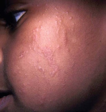
A higher clinical index of suspicion may be required to identify contact dermatitis, such as this, in children of color.
The condition can be present at birth. The macules and pustules can appear anywhere on the body, but most often on the chin, neck, upper chest, and/or lower back. The good news is they fade over time and are benign, so no treatment is necessary. The differential diagnosis from such conditions as herpes simplex and erythema toxicum is important, however, said Dr. Treadwell. One tip is to check for lesions on the palms and soles, which can be diagnostic in transient neonatal pustular melanosis, but not for erythema toxicum. A biopsy can confirm your clinical suspicions.
The cutaneous manifestations of neonatal lupus erythematosus can tip you off to this condition, she said. Skin lesions can be annular, discoid, or atrophic. Some children present with "raccoon eyes." Because this condition is related to maternal antibodies passed through the placenta during gestation, lesions generally clear by 6 months to 1 year of age.