Dermpath Diagnosis

Granular Cell Tumor

Author and Disclosure Information

Granular cell tumors (GCTs) tend to present as solitary nodules, not uncommonly affecting the dorsum of the tongue but also involving the skin, breasts, and internal organs. Cutaneous GCTs typically present as 0.5- to 3-cm firm nodules with a verrucous or eroded surface. They most commonly present in dark-skinned, middle-aged women but have been reported in all age groups and in both sexes. The differential diagnosis includes lepromatous leprosy, mastocytoma, reticulohistiocytoma, and xanthelasma.


References

Granular cell tumors (GCTs) tend to present as solitary nodules, not uncommonly affecting the dorsum of the tongue but also involving the skin, breasts, and internal organs.1 Cutaneous GCTs typically present as 0.5- to 3-cm firm nodules with a verrucous or eroded surface.2 They most commonly present in dark-skinned, middle-aged women but have been reported in all age groups and in both sexes.3 Multiple GCTs are reported in up to 25% of cases, rarely in association with LEOPARD syndrome (consisting of lentigines, electrocardiographic abnormalities, ocular hypertelorism, pulmonary stenosis, abnormalities of genitalia, retardation of growth, and deafness).4 Granular cell tumors generally are benign with a metastatic rate of approximately 3%.2

Granular cell tumors are histopathologically characterized by sheets of large polygonal cells with small, round, central nuclei; cytoplasm that is eosinophilic, coarse, and granular, as well as periodic acid–Schiff positive and diastase resistant; and distinct cytoplasmic membranes (Figure 1). Pustulo-ovoid bodies of Milian often generally appear as larger eosinophilic granules surrounded by a clear halo (Figure 2).5 Increased mitotic activity, a high nuclear-cytoplasmic ratio, pleomorphism, and necrosis suggest malignancy.6

Figure 1. Pseudoepitheliomatous hyperplasia and sheets of polygonal cells with light pink cytoplasm of a granular cell tumor (H&E, original magnification
×40).

Figure 2. Sheets of large polygonal cells with central nuclei; coarse, eosinophilic, granular cytoplasm; and large eosinophilic cytoplasmic pustulo-ovoid bodies of Milian characterize a granular cell tumor (H&E, original magnification ×600).

Lepromatous leprosy is characterized by sheets of histiocytes with vacuolated cytoplasm, some with clumped amphophilic bacilli known as globi (Figure 3). Mastocytoma can be distinguished from GCTs by the “fried egg” appearance of the mast cells (Figure 4). Although mast cells have a pale granular cytoplasm, they are smaller and lack pustulo-ovoid bodies and the polygonal shape of GCT cells. Reticulohistiocytoma, on the other hand, has two-toned dusty rose ground glass histiocytes (Figure 5), and xanthelasma can be distinguished histologically from GCT by the presence of a foamy rather than granular cytoplasm (Figure 6).

Figure 3. Lymphohistiocytic infiltrate with amphophilic collections of mycobacteria (globi) in lepromatous leprosy (H&E, original magnification ×600).


Figure 4.
Small “fried egg” mast cells with pale granular cytoplasm and admixed eosinophils in a mastocytoma (H&E, original magnification ×600).


Figure 5. Sea of histiocytes containing dusty rose or ground glass cytoplasm with two-toned darker and lighter areas in a reticulohistiocytoma (H&E, original magnification ×600).


Figure 6. Aggregates of histiocytes with foamy cytoplasm in xanthelasma (H&E, original magnifi-cation ×600).

Recommended Reading

Yellowish Papulonodular Periorbital Eruption
MDedge Dermatology
Subcutaneous Panniculitislike T-Cell Lymphoma
MDedge Dermatology
Granulomatous Changes Associated With Pigmented Purpuric Dermatosis
MDedge Dermatology
Pemphigus Vulgaris in Pregnancy
MDedge Dermatology
Lipidized Dermatofibroma
MDedge Dermatology
Hypopigmented Facial Papules on the Cheeks
MDedge Dermatology
Cutaneous Adenosquamous Carcinoma: A Rare Neoplasm With Biphasic Differentiation
MDedge Dermatology
Clear Cell Fibrous Papule
MDedge Dermatology
Sulfur Spring Dermatitis
MDedge Dermatology
Nodular Extramammary Paget Disease With Fibroepitheliomatous Hyperplasia
MDedge Dermatology

Related Articles

  • Dermpath Diagnosis

    Clear Cell Fibrous Papule

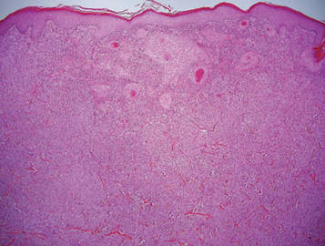
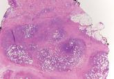
    A fibrous papule is a common benign lesion that usually presents in adults on the face, especially on the lower portion of the nose.
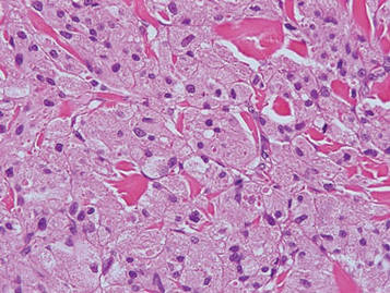
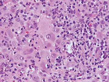
  • Dermpath Diagnosis

    Lipidized Dermatofibroma

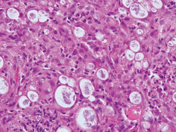
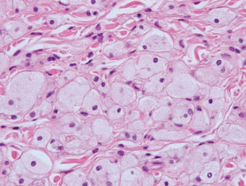
    Lipidized dermatofibromas most commonly are found on the ankles, which has led some authors to refer to these lesions as ankle-type fibrous...
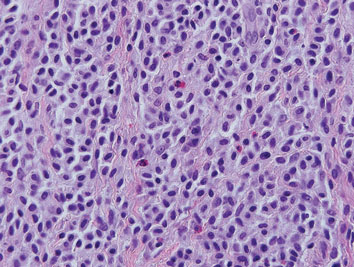