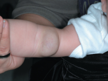
The FP recognized that the patient had a deep (cavernous) hemangioma. Hemangiomas consist of an abnormally dense group of dilated blood vessels. The proliferation phase occurs during the first year, and most growth takes place during the first 6 months of life. Proliferation then slows and the hemangioma begins to involute.
Hemangiomas may be superficial, deep, or a combination of both. Superficial hemangiomas (“strawberry” marks) are well-defined, bright red, and appear as nodules or plaques located on clinically normal skin. Deep (cavernous) hemangiomas (like this patient’s) are raised, flesh-colored nodules that often have a bluish hue and feel firm and rubbery. Most are clinically insignificant unless they impinge on vital structures, ulcerate, bleed, incite a consumptive coagulopathy, or cause high output cardiac failure or structural abnormalities.
In this case, there was no need for aggressive treatment because there were no signs of ulcerations or bleeding, and because the hemangioma was not blocking any of the infant’s essential organs. The FP reassured the parents that the hemangioma was likely to involute over time and might very well resolve completely without any treatment. The FP also explained that hemangiomas don't just pop open spontaneously—or secondary to minor trauma—and bleed. The parents were adequately reassured by this and planned to return for further follow up at the one-year well-child visit.
Photos and text for Photo Rounds Friday courtesy of Richard P. Usatine, MD. This case was adapted from: Usatine R, Madhukar M. Childhood hemangiomas and vascular malformations. In: Usatine R, Smith M, Mayeaux EJ, et al, eds. Color Atlas of Family Medicine. 2nd ed. New York, NY: McGraw-Hill; 2013:636-641.
To learn more about the Color Atlas of Family Medicine, see: www.amazon.com/Color-Family-Medicine-Richard-Usatine/dp/0071769641/
You can now get the second edition of the Color Atlas of Family Medicine as an app by clicking on this link: usatinemedia.com


