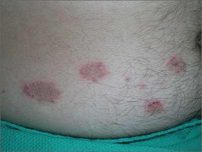
The FP concluded that this was a case of nummular eczema, after exploring other elements of the differential diagnosis, which included psoriasis, tinea corporis, and contact dermatitis. The FP looked at the patient's nails and scalp and saw no other signs of psoriasis. He then performed a potassium hydroxide (KOH) preparation and found no evidence of hyphae or fungal elements. (See a video on how to perform a KOH preparation here: http://www.mdedge.com/jfponline/article/100603/dermatology/koh-preparation.) He ruled out contact dermatitis because the history did not support it, and this wouldn’t have been a common location for it.
Nummular eczema is a type of eczema characterized by circular or oval-shaped scaling plaques with well-defined borders. (“Nummus” is Latin for “coin.”) Nummular eczema produces multiple lesions that are most commonly found on the dorsa of the hands, arms, and legs.
In this case, the FP offered to do a biopsy to confirm the clinical diagnosis, but noted that treatment could be started empirically and the biopsy could be reserved for the next visit only if the treatment was not successful. The patient preferred to start with topical treatment and hold off on the biopsy.
The FP prescribed 0.1% triamcinolone ointment to be applied twice daily. The FP knew that this was a mid-potency steroid and a high-potency steroid might work more rapidly and effectively, but the patient's insurance had a large deductible and the patient was going to have to pay for the medication out-of-pocket. Generic 0.1% triamcinolone is far less expensive than any of the high-potency steroids, including generic clobetasol.
Fortunately, at the one-month follow-up, the patient had improved by 95% and the only remaining problem was the hyperpigmentation, which was secondary to the inflammation. The FP suggested that the patient continue using the triamcinolone until there was a full resolution of the erythema, scaling, and itching. He explained that the post-inflammatory hyperpigmentation might take months (to a year) to resolve, and in some cases never resolves. The patient was not worried about this issue and was happy that his symptoms had improved.
Photos and text for Photo Rounds Friday courtesy of Richard P. Usatine, MD. This case was adapted from: Wah Y, Usatine R. Eczema. In: Usatine R, Smith M, Mayeaux EJ, et al, eds. Color Atlas of Family Medicine. 2nd ed. New York, NY: McGraw-Hill; 2013.
To learn more about the Color Atlas of Family Medicine, see: www.amazon.com/Color-Family-Medicine-Richard-Usatine/dp/0071769641/
You can now get the second edition of the Color Atlas of Family Medicine as an app by clicking on this link: usatinemedia.com


