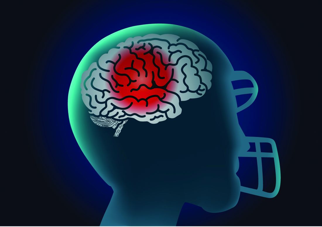
Imaging study provides detail, but not the full picture
Former athletes with a history of concussion averaged higher levels of total tau in their cerebrospinal fluid than did healthy controls, and those with the highest levels showed signs of reduced cognitive function in a case-control study.
 solar22/Thinkstock
Chronic traumatic encephalopathy (CTE) remains a postmortem diagnosis, but “the potential for treating postconcussion degeneration such as CTE depends on being able to detect the in vivo pathology at an early stage to intervene before the disease progresses to an irreversible stage,” wrote Foad Taghdiri, MD, of the University of Toronto and colleagues.
In a study published in Neurology, the researchers measured concentrations of phosphorylated tau181, total tau (t-tau), and beta-amyloid in the cerebrospinal fluid (CSF) of three groups: 22 former professional athletes who had suffered multiple concussions, 5 healthy controls, and 12 individuals diagnosed with Alzheimer’s disease (AD). The average ages of the groups were 56 years, 57 years, and 60 years, respectively. All the athletes were male, and their sports included snowboarding, hockey, and football.
The average t-tau level in the CSF of the athletes was significantly higher than that of controls (349.3 pg/mL vs. 188.8 pg/mL) and significantly lower than that of AD patients (857.0 pg/mL).
Normal CSF t-tau was defined as 300 pg/mL, and 12 former athletes (45%) had high t-tau levels, with an average of 499.3 pg/mL. In this group of high t-tau former athletes, the average score on the Trail Making Test (TMT) Part B was significantly lower than the average score among the 10 former athletes with normal CSF t-tau levels (t scores 45.6 vs. 62.3; P = .017).
In addition, results from MRI scans showed that fractional anisotropy values across all the tracts were significantly lower for those with high CSF t-tau levels, compared with those who had normal CSF t-tau levels (P = .036).
The findings were limited by several factors, including the small sample size, lack of female athletes, and limited ability to compare white matter integrity between high and normal CSF t-tau groups, the researchers noted.
However, the results suggest that “multiple concussive or subconcussive events may trigger neurodegeneration to a greater degree than expected on the basis of age alone,” they said. Although the study did not allow for diagnosing the participants with CTE, “we are engaged in longitudinal studies to track neurologic and neuropsychological function, CSF biomarkers, and structural brain changes over time to further assess the delayed effects of multiple concussions on the brain,” the researchers wrote.
The study was funded by the Toronto General and Western Hospital Foundation, PSI Foundation, and the Canadian Institute of Health Research. The researchers had no financial conflicts to disclose.
SOURCE: Taghdiri F et al. Neurology. 2019 May 8. doi: 10.1212/WNL.0000000000007608