Pediatric Dermatology Consult

A teen presents with a severe, tender rash on the extremities

A 13-year-old female is seen in urgent care for a 2-day history of severe tender rash on the arms and legs. The patient reported the rash started a few days after doing a long hike to a river with her grandfather. The day after the hike, her skin was very red and tender and as the day progressed, she started noticing some blisters on areas where she scratched the skin. The girl denied using any sunscreen or any other products on her skin on that day. The mom reported the younger brother also had some blisters on the arms but not as severe as the patient. She reported feeling hot and having severe pain on the skin. She has not developed any ocular or mucosal lesions.
She started taking lithium for depression and anxiety 3 weeks prior to her developing the rash. She denies taking any other medications, supplements, or recreational drugs.
She denied any prior history of photosensitivity, no history of mouth ulcers, joint pain, muscle weakness, hair loss, or any other symptoms.
Besides her brother, there are no other affected family members, and no history of immune bullous disorders or other skin conditions.
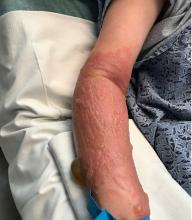
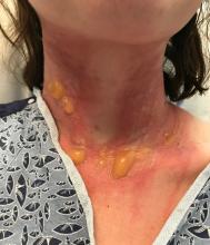
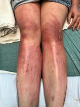
On physical exam, the girl appears in a lot of pain and is uncomfortable. The skin is red and hot, and there are tense bullae on the neck, arms, and legs. There are no ocular or mucosal lesions.

Your diagnosis is:

Bullous pemphigoid

Phyto photodermatitis

Pemphigus vulgaris

Bullous lupus erythematosus

Allergic contact dermatitis

“There’s rue for you, and here’s some for me; we may call it herb of grace o’ Sundays. O, you must wear your rue with a difference.”

— Ophelia in Hamlet by William Shakespeare

The patient was admitted to the hospital for IV fluids, pain control, and observation. The following day she admitted using the leaves of a plant on the trail as a bug repellent, as one time was taught by her grandfather. She rubbed some of the leaves on the brother as well. The grandfather shared some pictures of the bushes, and the plant was identified as Ruta graveolens.

Erythematous edematous patches with tense bullae.

Erythematous edematous patches with tense bullae.

The blisters were deroofed, cleaned with saline, and wrapped with triamcinolone ointment and petrolatum. The patient was also started on a prednisone taper and received analgesics for the severe pain.

Ruta graveolens also known as common rue or herb of grace, is an ornamental plant from the Rutaceae family. This plant is also used as a medicinal herb, condiment, and as an insect repellent. If ingested in large doses, it can cause severe abdominal pain and vomiting. It also can be hepatotoxic.

When applied to the skin and then exposed to the sun, it can cause severe phytophotodermatitis which can mimic a severe second-degree burn. The herb contains furocumarines, such as 8-methoxypsoralen and 5-methoxypsoralen and furoquinoline alkaloids. These chemicals when exposed to UVA radiation cause cell injury and inflammation of the skin. This is considered a phototoxic reaction of the skin, compared with allergic reactions, such as poison ivy dermatitis, which need a prior sensitization to the allergen for the T cells to be activated and cause injury in the skin. Other common plants and fruits that can cause phytophotodermatitis include citrus fruits, figs, carrots, celery, parsnips, parsley, and other wildflowers like hogweed.

Legs with linear erythematous patches and linear bullae

Legs with linear erythematous patches and linear bullae.

Depending on the degree of injury, the patients can be treated with topical corticosteroids, petrolatum wraps, and pain control. In severe cases like our patient, systemic prednisone may help stop the progression of the lesions and help with the inflammation. Skin hyperpigmentation after the initial injury may take months to clear, and some patient can develop scars.

The differential diagnosis should include severe bullous contact dermatitis like exposure to urushiol in poison ivy; second- and third-degree burns; severe medications reactions such Stevens-Johnson syndrome or toxic epidermal necrolysis, and inmunobullous diseases such as bullous lupus erythematosus, pemphigus vulgaris, or bullous pemphigoid. If there is no history of exposure or there are any other systemic symptoms, consider performing a skin biopsy of one of the lesions.

In this patient’s case, the history of exposure and skin findings helped the dermatologist on call make the right diagnosis.

Dr. Matiz is a pediatric dermatologist at Southern California Permanente Medical Group, San Diego. Email her at pdnews@mdedge.com.

References

J Burn Care Res. 2018 Oct 23;39(6):1064-6.

Dermatitis. 2007 Mar;18(1):52-5.

BMJ Case Rep. 2015 Dec 23;2015:bcr2015213388.

Recommended Reading

Hyperpigmented patch on the back
MDedge Family Medicine
Index finger plaque
MDedge Family Medicine
Study highlights differences between White and Latino patients with psoriasis
MDedge Family Medicine
Expert spotlights recent advances in the medical treatment of acne
MDedge Family Medicine
Don’t discount ultrapotent topical corticosteroids for vulvar lichen sclerosus
MDedge Family Medicine
Chronic, nonhealing leg ulcer
MDedge Family Medicine
A 31-year-old with a 3-week history of a waxing and waning, mildly pruritic eruption on his neck, chest, and back
MDedge Family Medicine
Neurofibromatosis type 1: More than skin deep
MDedge Family Medicine
Does early introduction of peanuts to an infant’s diet reduce the risk for peanut allergy?
MDedge Family Medicine
Rapidly developing vesicular eruption
MDedge Family Medicine