Evidence of diffuse axonal injury has been detected using an advanced form of MRI in military personnel who were clinically diagnosed as having only mild blast-related traumatic brain injury, according to a report in the June 2 issue of the New England Journal of Medicine.
This is the first time that widespread structural brain damage reflecting axonal injury has been identified in "mild" blast-related traumatic brain injury (TBI). In addition, the study confirms that axonal injury might be a primary feature of such wounds, as animal studies and computer simulations have suggested, said Christine L. MacDonald, Ph.D., of Washington University in St. Louis, and her associates.
The findings, however, cannot yet be considered definitive because all of the subjects in this study also sustained another mechanism of head injury simultaneously with the blast-related injury. All of them experienced additional head injury from a fall or motor vehicle crash, or from being struck by blunt objects at the time of the explosion, so the effects of the blast cannot be differentiated from the effects of these traumas, the investigators noted.
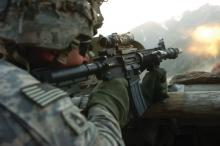
 Photo courtesy Wikimedia Commons/U.S. Army Sgt. Matthew C. Moeller/Public Domain
A U.S. Army soldier with 1st Battalion, 32nd Infantry Regiment, 10th Mountain Division, fires his weapon during a gun battle with insurgent forces in Barge Matal, Nuristan Province, Afghanistan, during Operation Mountain Fire on July 12, 2009. A study has found evidence that even "mild" blast-related traumatic brain injuries involve axonal damage that can be detected using diffusion tensor imaging.
They studied 84 members of the military who were injured in explosions in Iraq or Afghanistan during a 5-month period. A total of 63 had blast-related TBI that was judged on clinical assessment to be mild and uncomplicated, and 21 who had other injuries, chiefly orthopedic or soft-tissue wounds in the arms or legs, served as controls.
The study subjects underwent initial brain MRI when they were evacuated to Landstuhl Regional Medical Center in Germany immediately after they were wounded and follow-up MRI 6-12 months later at Washington University.
In addition to regular MRI, the subjects’ brains were scanned using diffusion tensor imaging (DTI), an advanced MRI method that measures water diffusion in many directions. Reduced anisotropy (directional asymmetry) of water diffusion on DTI is a marker of traumatic axonal injury.
Conventional MRI revealed abnormalities in only one of the TBI subjects, but DTI demonstrated reduced anisotropy in 18 (29%) of these subjects, across several of the 17 brain regions assessed in each subject. An additional 20 TBI subjects (32%) showed an abnormality within a single brain region on DTI.
"Quantitative analyses indicated significant reductions in relative anisotropy in the group of subjects with TBI as compared with the control group," Dr. MacDonald and her colleagues said (N. Engl. J. Med. 2011;364:2091-100).
Compared with control subjects, the TBI patients showed marked abnormalities in the middle cerebral peduncles, in cingulum bundles, and in the right orbitofrontal white matter – areas that have been predicted by computer simulations to sustain the most intense stresses during a blast. These are not the same regions of the brain that are commonly injured in civilian cases of TBI that do not involve blast exposure, they noted.
"The characteristics of the abnormal DTI signals changed between initial scanning and follow-up scanning, in a fashion that was consistent with the evolution of relatively acute injuries," the researchers said.
The follow-up scans indicated that the edema and cellular inflammation accompanying the axonal injury was resolving while the axonal injury itself persisted.
The investigators said that DTI assessments "may be useful in diagnosis, triage, and treatment planning in clinical practice" since the method "can be performed relatively quickly on the MRI scanners at U.S. military facilities and civilian hospitals."
Dr. MacDonald and her associates emphasized, however, that the findings in this study are preliminary and that TBI remains primarily a clinical diagnosis. "Normal findings on a DTI scan do not rule out TBI, nor are DTI findings in isolation sufficient to make this diagnosis with certainty," they noted.
The investigators noted several limitations of the study, including a moderate sample size and an all-male study population. Several studies of the relationship between DTI abnormalities and clinical outcomes are currently under way, they added.
This study was supported by the Congressionally Directed Medical Research Program and the National Institutes of Health. Dr. MacDonald’s associate, Dr. David L. Brody, reported ties to Wuestling and James Medicolegal Consulting, Burroughs Wellcome Fund, Thrasher Research Fund, the Hope Center for Neurological Disorders, the International Neurotrauma Society, and the Defense and Veterans Brain Injury Center. Dr. Ropper reported no financial conflicts of interest relevant to his commentary.

