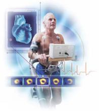
Order exercise stress testing without imaging for patients with a low to intermediate probability of coronary artery disease (CAD), unless preexisting electrocardiographic (EKG) changes would render such a test nondiagnostic. C
Order stress testing with imaging for patients with preexisting EKG changes and/or a high probability of CAD. C
Do not use stress testing to screen asymptomatic patients for CAD. C
Consider pharmacologic testing for patients who are unable to exercise to an appropriate cardiac workload; it has the same predictive value as a nuclear exercise stress test. B
Strength of recommendation (SOR)
A Good-quality patient-oriented evidence
B Inconsistent or limited-quality patient-oriented evidence
C Consensus, usual practice, opinion, disease-oriented evidence, case series
Exercise has been used for cardiac stress testing for decades. But testing and imaging techniques and knowledge of the efficacy of this common diagnostic tool continue to evolve. Optimizing your use of stress testing requires that you familiarize yourself with the latest evidence. The evidence-based answers to these 6 questions will help you do just that.
1. How reliable are exercise stress tests?
That depends, of course, on any number of variables, including the protocol utilized, the number of stenotic vessels, the degree of stenosis, and even the sex of the patient.
False-negative and false-positive results are frequent in treadmill testing without imaging. (For more on the different protocols, see “Standard and nuclear exercise stress tests: A look at protocols”) Sensitivity is related to the number of stenotic vessels and the degree of stenosis. For a man with single-vessel disease and ≥70% stenosis, the likelihood of an abnormal test is only 50% to 60%. Even in a man with left main artery disease, the sensitivity is only about 85%.1
In some cases, failure to reach a cardiac workload sufficient to produce ischemia can lead to a false-negative test, and it is up to the physician performing the test to label it as nondiagnostic. Other reasons for false-positive or false-negative results include preexisting ST segment abnormalities, which can cause false-positive elevation of the ST segment during exercise; the use of digitalis, which affects the ST segment; and the presence of ventricular hypertrophy or cardiomyopathy.1 Patients with any of these conditions should undergo stress testing with imaging instead.
Nuclear stress testing is indicated for patients who have baseline EKG abnormalities, suspected false-positive or false-negative results from a stress test without imaging, known CAD or previous revascularization, a pacemaker, or a moderate to high likelihood of a CAD diagnosis. The addition of a tracer isotope and imaging boosts the test’s predictive value.1
The positive predictive value of nuclear stress testing is difficult to calculate because an abnormal test should lead to initiation of therapy designed to reduce the risk of cardiac death or myocardial infarction (MI). Numerous studies have found the rate of cardiac events after a negative radionuclide stress test to be less than 1% per year.2 The event rate after a negative test is lower in women than in men; after a positive test, however, the event rate in women is 2 to 3 times higher.2,3 Overall, stress testing is less sensitive in women than in men, at least in part because of their lower likelihood of CAD associated with any given symptom set.4
Exercise stress testing can be done with a number of treadmill protocols. The most widely used are:18,19
- the Bruce Protocol (the most common),1 which increases the slope of the treadmill and the speed of the belt in 3-minute intervals;
- the modified Bruce Protocol, a less aggressive format in which slope and speed are alternatively increased; and
- the naughton Protocol (typically reserved for patients whose ability to walk is limited), which starts with a very slow belt speed and a nearly flat slope and increases both elements slowly.
During the test, heart rate and BP are measured, along with continuous EKG monitoring, but the frequency of BP measurement and 12-lead EKG printouts varies among testing facilities.
Patients must attain a heart rate of 85% of their age-predicted maximum for the test to be considered diagnostic; they typically exercise until they’re unable to continue or they develop symptoms that prompt the clinician performing the test to stop it. Monitoring continues for some time after the patient stops exercising—usually 4 to 5 minutes in an asymptomatic patient, or until any symptoms and EKG changes that developed during the test resolve. If chest pain or EKG changes persist, the patient may need to be admitted to the hospital.
The procedure for nuclear stress testing is similar, except that the patient must estimate when he or she can only walk for 1 more minute. A tracer isotope is injected at that time.
For years, thallium was used for this purpose. However, thallium is taken up by the perfused myocardium and has the drawback of rapid redistribution with resolution of ischemia, which can lead to false-negative tests.20
Technetium (99mTc-labeled sestamibi), which is commonly used for other nuclide scans, is now the preferred isotope for nuclear stress tests.21 It is taken up by mitochondria in the perfused myocardium and does not redistribute, which results in fewer false-negative scans. Additionally, the energy emitted by 99mTc-labeled sestamibi is higher and produces cleaner pictures.21
Single photon emission computed tomography (SPECT) scans are taken in 3 planes as part of the nuclear stress test. A set of resting scans is taken before the exercise test. The isotope is then allowed to wash out and another dose is injected at peak cardiac workload so a second set of scans can be taken and compared with the resting images.
Perfusion defects that are present both at rest and with stress indicate an area of infarction, whereas defects that appear with stress but not at rest indicate ischemia. The probable location of the coronary artery lesions responsible for the ischemia can be inferred from the area in which the defects appear.
Gated imaging—serial images that are coupled with EKG changes, then reassembled to produce a moving image of the heart—is now usually part of the process. The result can be examined for areas of wall motion abnormalities and used to calculate an ejection fraction.22


