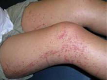
A mother brings her 5-year-old boy in to your office because she is concerned about a rash on his legs that seems to be worsening. She tells you that he had a runny nose and a mild cough a week earlier, but that those symptoms resolved before the rash developed. He has also complained of “belly pain.”
The boy’s mother says he’s been less active and more irritable since the onset of the rash, and that he is hardly eating. She also tells you that earlier in the day, her son told her that it hurts to walk.
You dig deeper…
A complete review of systems is otherwise negative. The 5-year-old was born at term without complication. He has met all developmental milestones and his immunizations are up to date. He takes no medications.
The boy’s vital signs are normal. He has an erythematous maculopapular rash distributed on his legs symmetrically; it is palpable, nontender, and nonblanching. You detect no abnormalities in abdominal, neurologic, or musculoskeletal examinations.
A complete blood count (CBC) and basic metabolic panel (BMP) reveal mild leukocytosis with a normal differential. Urinalysis shows moderate blood and trace protein. Laboratory results are otherwise normal.
WHAT IS YOUR DIAGNOSIS?
A classic presentation revealed: Henoch-Schönlein purpura
The combination of rash (FIGURE), abdominal pain, arthralgia, and evidence of renal involvement are the classic symptoms for Henoch-Schönlein purpura (HSP),1 the most common systemic vasculitis of childhood.2 Fever may also be present. Boys are affected nearly twice as often as girls. HSP occurs only rarely among adults, and men and women are affected equally. The estimated annual incidence in children is 20 cases per 100,000, with most cases occurring in those between the ages of 4 and 6 years.3
Although the exact cause of HSP is unknown, 75% of cases follow an upper respiratory infection. Streptococcus, Mycoplasma, adenovirus, parvovirus, Epstein-Barr virus, and varicella have all been implicated as offending pathogens. A number of case reports have also described the disease after the use of certain drugs, including penicillin, ampicillin, and quinine; and after the administration of vaccines, including those for typhoid, measles, yellow fever, and cholera. In all cases, the underlying mechanism is thought to be a systemic rise in immunoglobulin A (IgA), which forms immune complexes deposited in arterioles, capillaries, and venules.4 The precise interactions in the mechanism of disease are yet to be determined.
FIGURE
Rash and leg pain
Like the patient described in the text, this 11-year-old girl had a similar rash on her legs, as well as leg and abdominal pain.
HSP is usually benign, self-limited
HSP signs and symptoms may occur in any order over a period of days to weeks. Symptoms tend to be less severe in younger children than in older children and adults. Besides abdominal and renal systems, the lungs and central nervous system (CNS) may be affected, although rarely in children. Pulmonary involvement, if it does occur, usually manifests as diffuse alveolar hemorrhage or interstitial pneumonia or fibrosis.5 CNS vasculitis may lead to cerebral hemorrhage.6
HSP usually is a benign, self-limited disorder. The average disease course is 4 weeks, although it may be as brief as 3 days or as long as 2 years. Up to 94% of children (and 89% of adults) recover fully.7 However, the potential for chronic renal disease does exist.8
The recurrence rate of HSP is generally reported as 40%;8 patients who develop nephritis have higher recurrence rates.9
Rash is almost always present
Nearly all of those affected with HSP exhibit a rash. In children, it may begin as urticarial or maculopapular skin lesions. The rash typically is a palpable purpura, with lesions measuring 2 to 10 mm in diameter. Classically, the rash appears symmetrically on the extensor surfaces of the arms, legs, and buttocks. Lesions may also occur on the face and ears; however, the rash usually spares the trunk.2 The rash generally fades over several days and gives way to darkened pigment. The purpuric lesions resolve more quickly with bed rest and tend to reappear when the patient resumes activity.
Bullous or necrotic lesions—although reported to occur in up to 60% of affected adults—are uncommon in children. If you had noted such lesions in this case, you would have had to rule out toxic vasculitides and meningococcal septicemia or other septic emboli.10
Arthralgia affects lower extremities
Arthralgia is the second most common clinical manifestation of HSP, occurring in approximately 82% of patients. Joint pain associated with HSP is likely due to periarticular soft tissue edema, and most commonly occurs in the hips, knees, or ankles.1,11 The arthralgia of HSP is transient and self-limited and does not cause permanent damage to the joints. Like the rash, joint pain tends to decrease with bed rest and increase with activity.11


