A 60-YEAR-OLD WOMAN came into our primary care clinic and asked that we look at a rash on her left shin. The rash started as a few small red spots 8 months earlier that gradually merged and turned into one shiny irregular area with brownish discoloration. The rash was neither itchy nor painful, and remained unchanged during exposure to sunlight.
The patient indicated that she had been diagnosed with type 1 diabetes when she was 24 years old and was using a continuous subcutaneous insulin infusion device. She said that over the years she’d developed diabetic retinopathy, polyneuropathy, and end-stage renal disease requiring hemodialysis.
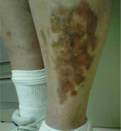
Clinical exam revealed an 8 × 6 cm well-demarcated red-brown irregular plaque on her left shin. The lesion had a yellow atrophic center and extensive telangiectasias (FIGURE). There were no similar lesions anywhere else on her body.
FIGURE
Red-brown plaque over left shin with yellow atrophic center and extensive telangiectasias

WHAT IS YOUR DIAGNOSIS?
HOW WOULD YOU TREAT THIS PATIENT?

