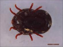
During my preceptorship, I (PK) encountered a 67-yearold cattle rancher with a month-long history of right ear pain, right-sided headaches, hearing loss, and occasional dizziness. He’d seen 2 other physicians on separate occasions who had prescribed antibiotics and ear drops for cerumen removal, yet his symptoms persisted. A computed tomography (CT) scan was normal.
When I examined the patient, his right inner ear canal showed a white, crusting exudate condensed in the tympanic membrane area. I inserted the otoscope farther into the canal and observed a single insect leg sticking out from the grey mass. A resident used the otoscope and forceps to extract the live specimen intact. It was identified as an Otobius tick.
Despite having a tick in his ear canal for more than a month, the patient was doing well at his 2-week follow-up appointment and showed no signs of tick-borne illness. The appearance of the tick had closely resembled impacted cerumen, which had led to delayed diagnosis and an unnecessary CT scan.
A careful otic exam was paramount, because directly viewing the insect’s extremity was the key to diagnosis.
Petra Kelsey, 2nd year medical student
Marfa, Texas
Adrian Billings, MD, PhD
Galveston, Texas


