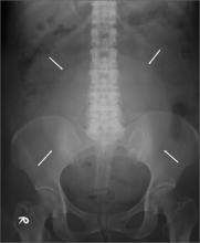
This patient had a large uterine fibroid. Also known as a leiomyoma, a uterine fibroid is a benign tumor that is composed of smooth muscle tissue and fibrous connective tissue. It is the most common pelvic tumor found in the female body. Unlike cancerous tumors, fibroids usually grow slowly and do not break away or invade other parts of the body.
Based on its position within the uterus, a fibroid can be submucosal, intramural, or subserosal. Uterine fibroids are usually diagnosed based on a clinical history and pelvic examination; the presence of a fibroid is confirmed by ultrasound, magnetic resonance imaging, computed tomography, saline infusion sonography, or hysterosalpingography.
Small fibroids that are asymptomatic or cause only minor problems are not usually treated. However, if a fibroid is large or results in pain or excessive bleeding, further management may be needed. Management of fibroids may be surgical or nonsurgical (hormones). Factors that affect management choices include the patient’s desire to become pregnant or preserve her uterus, symptom severity, and tumor characteristics.
This patient’s physicians originally suspected malignancy, so they consulted a gynecologist for a sonographic examination. The ultrasound revealed a heterogeneous mass with calcification. The patient underwent a hysterectomy.
The solitary soft tissue mass that was removed measured 13 cm in diameter and weighted 2100 g. Histopathological analysis revealed that the fibroid was made up of myometrium and fibrous connective tissue. After hospital discharge, the patient resumed all of her normal activities with no recurrence.
Adapted from: Chang CF, Ke CY, Lin JG. Photo Rounds: large pelvic mass in a 46-year-old woman. J Fam Pract. 2013;62:581-584.


