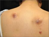
Based on his clinical presentation and skin biopsy results, the patient was given a diagnosis of cutaneous sarcoidosis. A biopsy from the right side of his nose demonstrated sarcoidal granulomas. Acid-fast bacilli and periodic acid-Schiff stains were negative. A biopsy of one of the tattoo nodules showed sarcoidal granulomas, and close inspection revealed red tattoo pigment within the granulomatous inflammation.
X-rays showed bilateral hilar lymphadenopathy, which was consistent with pulmonary sarcoidosis, and the lace-like appearance of the middle and distal phalanges was consistent with skeletal sarcoidosis.
Systemic sarcoidosis is an idiopathic, granulomatous disease that affects multiple organ systems but primarily the lungs and lymphatic system.1 The estimated prevalence of systemic sarcoidosis ranges from less than 1 to 40 cases per 100,000 people, and the condition is more common among African Americans.1
Cutaneous sarcoidosis can occur as a manifestation of systemic sarcoidosis. It occurs in 20% to 35% of patients with systemic sarcoidosis2 and may present as asymptomatic red or skin-colored papules and firm nodules within tattoos, old scars, or permanent makeup. Cutaneous sarcoidosis in tattoos may be the first manifestation of sarcoidosis, and the time between acquiring the tattoo and developing sarcoidal nodules varies widely.
It is not clear why sarcoidal granulomas occur in tattoos. One possibility is that chronic low-grade exposure of the immune system to foreign materials such as tattoo ink leads to granulomatous hypersensitivity.2,3 Sarcoidosis usually occurs in red (cinnabar), black (ferric oxide), or blue-black areas of tattoos,4 in which the pigment acts as a nidus for granuloma formation.
A skin biopsy is helpful in making a diagnosis of cutaneous sarcoidosis. The histopathology shows noncaseating epithelioid granulomas.
Because skin manifestations may be the first and only sign of systemic sarcoidosis, patients with cutaneous sarcoidosis should be evaluated for systemic disease. Cutaneous sarcoidosis has been associated with bilateral hilar lymphadenopathy, pulmonary sarcoidosis, uveitis, arthritis, and dactylitis.3
