Photo Rounds

Pleuritic chest pain and globus pharyngeus

Author and Disclosure Information

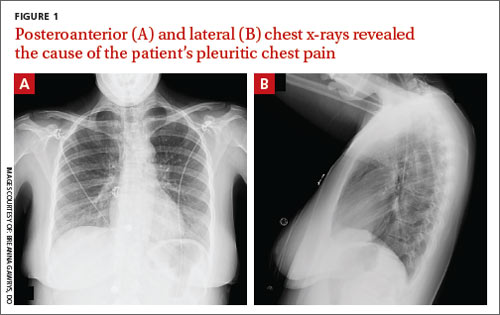
An AP chest x-ray provided no clues as to the cause of the patient’s substernal chest pain. Two other x-ray views, however, made the diagnosis clear.


References

A 22-year-old woman with a history of attention-deficit/hyperactivity disorder and childhood asthma came to the emergency department (ED) for treatment of a cramping, substernal, pleuritic chest pain she’d had for a week and the feeling of a “lump in her throat” that made it difficult and painful for her to swallow. The patient’s vital signs were normal and her substernal chest pain was reproducible with palpation. An anteroposterior (AP) chest x-ray (CXR) was unremarkable.

A “GI cocktail” (lidocaine, Mylanta and Donnatal), ketorolac, morphine, and lorazepam were administered in the ED, but did not provide the patient with any relief. She was admitted to the hospital to rule out acute coronary syndrome and was kept NPO overnight. A repeat CXR with posteroanterior (PA) and lateral views was also obtained (FIGURE 1A AND 1B).

WHAT IS YOUR DIAGNOSIS?
HOW WOULD YOU TREAT THIS PATIENT?

Pages

Recommended Reading

Epinephrine use for anaphylaxis in schools: First national survey
MDedge Family Medicine
Beware common management pitfalls in severe refractory pediatric AD
MDedge Family Medicine
SLIT: Guidelines in progress and practical concerns
MDedge Family Medicine
When to use SLIT and SCID in atopic dermatitis
MDedge Family Medicine
VIDEO: Novel three-shot immunotherapy regimen
MDedge Family Medicine
Pimecrolimus cream safe, effective for atopic dermatitis in young children
MDedge Family Medicine
Early pneumonia linked with later asthma and COPD
MDedge Family Medicine
Aspirin desensitization making headway in U.S.
MDedge Family Medicine
Send kids home 2 hours after food challenge testing
MDedge Family Medicine
AAAAI: Grass allergy tablets show favorable NNT
MDedge Family Medicine