ROME – Starting cardiac rehabilitation sessions within 2 weeks of an acute myocardial infarction is safe and can improve cardiac perfusion and function, according to results of a randomized, controlled study of 46 patients who had residual myocardial ischemia following an MI and percutaneous coronary revascularization.
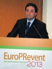
"Exercise-induced changes in myocardial perfusion and function were associated with an absence of unfavorable left-ventricular remodeling and an improved cardiovascular functional capacity," Dr. Francesco Giallauria said at the European Association for Cardiovascular Prevention and Rehabilitation* annual meeting. He hypothesized that exercise training begun soon after an MI improves myocardial perfusion by inducing coronary vascular adaptation or enhancing development of collateral cardiac vessels.
The study began with patients who underwent percutaneous coronary intervention (PCI) following an acute ST-elevation MI who were then assessed by dipyridamole stress and gated single-photon emission CT (SPECT) using labeled sestamibi. All 46 included patients had a significant amount of residual myocardial ischemia despite PCI, presumably caused by inadequate perfusion in myocardial regions not vascularized by the PCI-treated coronaries, said Dr. Giallauria, a cardiologist at the University of Naples (Italy). All patients also underwent a cardiopulmonary exercise test at baseline to determine their peak exercise capacity.
The researchers then randomized the patients. Twenty-five entered a hospital outpatient program of supervised, 30-minutes sessions of bicycle exercise three times a week, with an exercise target during each session of 60%-70% of their peak exercise capacity. The program began within 2 weeks of their acute MI. The remaining 21 patients served as controls and received just instructions about maintaining physical exercise and lifestyle modification.
The exercise rehabilitation program continued for 6 months, when all 46 patients underwent a follow-up examination of their myocardial perfusion with gated SPECT and follow-up cardiopulmonary stress testing. The average age of the patients was 54 years, and 87% were men.
The results showed that after 6 months, patients who underwent exercise training had an average 29% increase in peak exercise capacity and a 10% average rise in their peak heart rate compared with baseline, both statistically significant differences. Results from the serial SPECT studies showed that left ventricular ejection fraction rose from an average of 48% at baseline to 52% in the patients who underwent exercise training, a statistically significant rise, and their wall motion and wall thickness scores showed improvements in cardiac shape and function compared with baseline, all differences that were statistically significant. In contrast, the control patients had no significant changes for any of these exercise or cardiac measures during the 6 months of follow-up.
In addition, the SPECT imaging showed that at follow-up the extent of myocardial perfusion defect and ischemia fell by about 50% in the exercise group while remaining virtually unchanged in the control patients.
Dr. Giallauria said that he had no disclosures.
On Twitter @mitchelzoler
*Correction, 5/29/2013: An earlier version of this story misstated the name of the European Association for Cardiovascular Prevention and Rehabilitation annual conference.


