The clinical picture:
- Sharp chest pain radiating down the right arm
- Syncope
- Temp 97.3°F, pulse 78, respirations 21, BP 162/96 mm Hg, O2 saturation 98% on room air
- ECG: normal sinus rhythm, rate 70, and no ST-T wave changes
A 47-year-old African American woman came into our emergency department with right-sided chest pain that she described as sharp, and radiating down her right arm. She said she’d lost consciousness for a few seconds with only minimal exertion. She told us that she was being treated for hypertension by her primary care physician, and indicated that she’d never had a fainting spell before.
Our patient’s vital signs were:
- temperature 97.3°F
- pulse 78 beats per minute, respirations 21
- blood pressure 162/96 mm Hg
- O2 saturation 98% on room air.
She had a regular heart rate and rhythm, no murmurs, no rubs, no gal lops, and her pulses were 2+ bilateral and equal.
Her lung exam was clear to auscultation bilaterally; she had no tachypnea, wheezes, or crackles. The remainder of her physical exam was unremarkable. She was taking verapamil once a day and Excedrin as needed, but no other medications.
Our patient’s electrocardiogram (ECG) showed a normal sinus rhythm, rate 70, and no ST-T wave changes. We admitted her for further evaluation.
Following serial cardiac enzymes and ECGs, we ruled out a myocardial infarction. We ordered an exercise stress test, but it had to be stopped when she developed light-headedness and hypotension.
What tests would you order next?
What do you suspect is causing this woman’s chest pain and fainting spells?
What do you suspect is causing this woman’s chest pain and fainting spells?
Echo unlocks the mystery
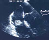
We ordered a transesophageal echo- cardiogram, which revealed a spherical 2 cm x 1.5 cm mass attached to the lateral wall of the right atrium just above the tricuspid valve (FIGURE, TOP LEFT). Differential diagnoses for this mass were atrial myxoma, intracardiac thrombus, lipomatous infiltrations, and calcified mitral annulus.1 A positron-emission tomography (PET) scan was nondiagnostic.
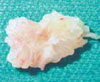
The mass was surgically excised. Pathology showed right atrial papillary myxoma, which was negative for signs of malignancy (FIGURE, BOTTOM LEFT).
A rare cardiac mass
While rare, papillary myxomas are the most common primary cardiac mass. On histologic exam, they are gelatinous structures that consist of myxoma cells embedded in a stroma rich in glycosamino- glycans.2,3 On macroscopic exam, myxomas are pedunculated and have a variable surface. These masses produce vascular endothelial growth factor, which could contribute to the malignant potential of histologically benign myxomas.4,5
Listen for “tumor plop”
Symptoms of myxomas depend on the location of the tumor. Myxomas are frequently accompanied by constitutional symptoms such as fever and weight loss (although our patient had neither). Embolization is also associated with such lesions.
Left-sided lesions present with symptoms of mitral valve obstruction, along with electrocardiographic changes of left atrial hypertrophy.2 Ventricular myxomas may cause outflow obstruction.3
The signs and symptoms of myxoma may have a sudden onset or be positional in nature, reflecting changes in tumor position due to gravity. An auscultatory finding, called “tumor plop,” is a characteristic low-pitched sound that may be audible during early or mid-diastole,6 though we heard no such sound during our evaluation of our patient.
Our patient was unusual
Diagnosis is made by echocardiography, and most (80%) of myxomas are found in the left atrium. Atrial myxoma typically appears as a well-circumscribed mobile mass that is attached to the atrial septum. Our patient, however, had a myxoma located in the right atrium that was attached to the lateral wall. Definitive diagnosis is made by surgical excision.
Treatment: Prompt resection
Due to the risk of embolization and cardiovascular compromise, prompt resection is recommended and is generally curative, as it was for our patient.3,7,8 Postoperative mortality rate after resection is under 5%. For multiple recurrent tumors, cardiac transplantation should be considered.1,9
Correspondence
Family medicine Department, John Peter Smith Hospital, 1500 South main St, Fort Worth, TX 76104; hrana@ jpshealth.org