CHICAGO – For periorificial dermatitis, don’t do a thing. That was the first-line therapy suggested at this year’s American Academy of Dermatology summer meeting.
"Sometimes you have to be brave to tell a patient you’re sending them home with nothing," said Dr. Sarah Wolfe of Duke University in Durham, N.C. "But ‘zero’ therapy is important. It’s simply counseling your patient to recognize the disease and avoid anything that could be exacerbating or precipitating it."
In theory, this advice includes abstinence from using any cosmetics for about 2 months, but "I am not sure whom that will work for," said Dr. Wolfe.
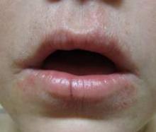
Periorificial dermatitis can affect anyone, but typically occurs in women aged 20-45 years across all ethnicities; it generally presents as erythematous pustules up to 2 mm in size near the nose and mouth, and patients report a burning sensation.
In children, the condition occurs equally in both genders, and tends to peak by age 5 years. Some children develop a distinctive form of the disease – childhood granulomatous periorificial dermatitis – that features persistent, inflammatory papules and pustules that appear symmetrically around the mouth. Although this form tends to be harder to treat and lasts longer, reassure patients that the condition will heal without scarring, Dr. Wolfe said.
Role of steroids
The minimalist approach to treating periorificial dermatitis is particularly important if the condition is related to steroid use, Dr. Wolfe said. Although the prevailing wisdom has been that corticosteroids caused periorificial dermatitis, enough evidence exists to show that corticosteroids likely exacerbate, rather than cause the problem, she explained.
Sometimes patients will use topical steroids (often medications prescribed for other family members or for other ailments) to help calm the burning, unaware of steroids’ potential to make their symptoms worse, she added.
If steroids are implicated, prepare your patient for the possibility of a steroid rebound that lasts a couple of weeks once they stop using the topicals.
"You have to either counsel them through it, or consider using a lower-potency steroid such as a hydrocortisone 2.5% or 1% to get them through that period," Dr. Wolfe said.
Inhaled steroids also have been associated with periorificial dermatitis, occurring where the inhalant mask touches the skin and lips. Intranasal steroids, particularly mometasone furoate and fluticasone furoate, have similarly been associated with the condition in cases of pustules starting near the nose and then spreading to the mouth and chin (Allergy 2001;56:944-8), (Cutan. Ocul. Toxicol. 2012;31:160-3).
Systemic steroids such as prednisone have been associated with the development of periorificial dermatitis upon tapering them, although this association has not been well studied. Because of the risk for the unintentional transfer of topical steroids to the face from other parts of the body being treated, counsel patients to be scrupulous about handwashing after applying their medications, Dr. Wolf emphasized.
Similarly, unintentional corticosteroid use can occur with the use of over the counter skin-lightening products marketed as "herbal" or "all natural," she said.
Oral, topical options
If you and your patient prefer to actively treat the condition, Dr. Wolfe recommended using the most evidence-based therapy available: Oral tetracycline 250-500 mg twice per day, a therapy which showed clearance in 4 to 8 weeks when compared to placebo (G. Ital. Dermatol. Venereol. 2010 Aug;145:433-44).
She did not recommend using this therapy in children younger than 8 years, because of the drug’s association with dental abnormalities. Other antibiotics such as doxycycline, minocycline, and azithromycin (in children) have not been well studied, Dr. Wolfe said, although she noted anecdotal reports of pediatric dermatologists prescribing azithromycin if the patient’s presentation is "slightly disfiguring."
Although erythromycin may be easier to use when controlling costs, both it and pimecrolimus are viable options for topical therapy, Dr. Wolfe said. Pimecrolimus 1% cream has been shown to reduce disease severity in 2 weeks, while erythromycin 2% solution, in ointment or gel form twice daily, brought clearance in 3-7 weeks in a randomized, placebo-controlled trial. She emphasized the importance of discussing all topical exposures with patients, regardless of the treatment option chosen.
Dr. Wolfe had no financial conflicts to disclose.
On Twitter @whitneymcknight


